

Neurofilament impairment in neurodegenerative diseases: pathogenesis, biomarkers and diagnostic values
 0
0 Abstract
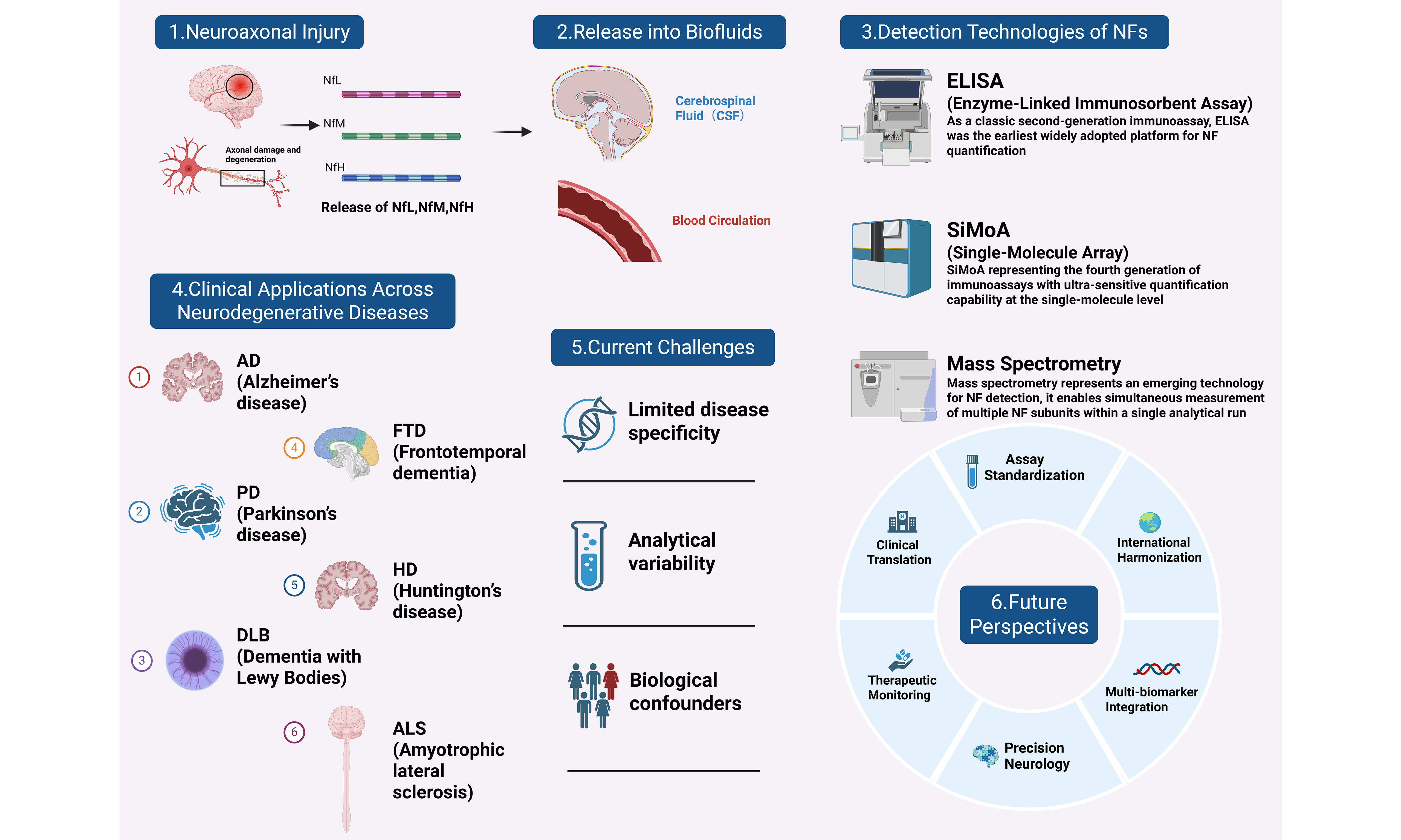
Neurofilament (NF) proteins, as integral components of the neuronal cytoskeleton, are essential for maintaining axonal structure and function. Genetic defects, aberrant post-translational modifications, and co-aggregation with other pathogenic proteins can disrupt NF homeostasis, leading to axonal injury and neuronal dysfunction. Advances in ultrasensitive detection technologies, particularly SiMoA assays, have enabled reliable quantification of NFs in both cerebrospinal fluid (CSF) and peripheral blood, substantially accelerating their clinical translation. However, current research has predominantly focused on the neurofilament light chain (NfL), with comparatively limited attention given to neurofilament medium (NfM) and heavy (NfH) chains. In this review, we comprehensively synthesize recent research findings in NfL, NfM, and NfH across major neurodegenerative diseases, including Alzheimer’s disease (AD), Parkinson’s disease (PD), dementia with Lewy bodies (DLB), frontotemporal dementia (FTD), Huntington’s disease (HD), and amyotrophic lateral sclerosis (ALS). Further, we provide a multi-level analysis spanning molecular structure, pathological mechanisms, and clinical translation, while comprehensively evaluating key confounding factors and methodological challenges that influence detection outcomes. We propose that future efforts should prioritize the universal standardization of detection methods and highlight the value in precision management of neurodegenerative diseases.
Keywords
INTRODUCTION
Neurodegenerative diseases have imposed a rapidly escalating burden on global public health. The limited healthcare resources and insufficient insurance coverage, particularly in low- and middle-income countries, frequently lead to the diagnosis of neurodegenerative diseases at advanced stages[1]. The lack of objective and quantifiable biomarkers for disease progression remains a major obstacle to effective clinical management and therapeutic development.
Alzheimer’s disease (AD) and Parkinson’s disease (PD) are the two most common neurodegenerative disorders[2]. The pathogenesis of AD is primarily characterized by extracellular β-amyloid (Aβ) deposition and intracellular accumulation of hyperphosphorylated tau, leading to synaptic dysfunction and neuronal degeneration[3]. In contrast, PD is mainly hallmarked by degeneration of dopaminergic neurons in the substantia nigra and abnormal aggregation of α-synuclein (α-syn), which subsequently results in both motor and non-motor symptoms[4]. The diagnosis of AD and PD typically relies on clinical assessment, neuroimaging, and biochemical evaluation. In clinical assessment, AD is primarily evaluated through cognitive function testing[5], whereas PD is mainly assessed based on motor symptoms[6]. Neuroimaging techniques such as magnetic resonance imaging (MRI) and positron emission tomography are used to detect brain atrophy and functional abnormalities[7]. Regarding biomarkers, cerebrospinal fluid (CSF) Aβ and tau proteins are widely used in AD research[8], while α-syn and dopamine-related indicators are commonly applied in PD[9]. However, current diagnostic approaches for AD, PD and other neurodegenerative diseases still have several limitations, including invasiveness, high cost, and insufficient sensitivity for early diagnosis. Therefore, there remains a need to develop reliable and minimally invasive biomarkers[10]. Emerging blood-based biomarkers, such as neurofilaments (NFs), have demonstrated considerable promise as indicators of axonal damage and disease progression[11].
NFs are cytoskeletal protein components of neurons and are divided into neurofilament light chain (NfL), medium chain (NfM), and heavy chain (NfH)[12]. Research on NFs dates back to silver-staining observations in neuroanatomy in the late nineteenth century[13]. Earlier studies, primarily based on CSF (human or animals) analyses and animal models, characterized the structural and functional properties of NFs. The C-terminal tail domains of NfM and NfH are longer and contain numerous phosphorylation sites, whereas the tail of NfL is shorter and exhibits a lower degree of phosphorylation[14]. Consequently, research progress on NfM and NfH has been relatively slow due to several factors, including immature detection methods, large molecular sizes, and complex phosphorylation states[15]. In contrast, benefiting from its smaller molecular weight, high solubility, and ready release into CSF and blood, NfL has been detected at significantly elevated levels in various neurodegenerative diseases using currently available technologies, establishing it as the predominant NF subunit[16].
In recent years, a major breakthrough has been the advent of ultra-high-sensitivity immunoassays, particularly single-molecule array (SiMoA) technology, which enables reliable and reproducible quantification of NfL in serum and plasma[17]. This transition is largely reflected in the expanding body of evidence supporting the diagnostic and prognostic utility of blood NfL in multiple neurodegenerative diseases, including AD, PD, and dementia with Lewy bodies (DLB)[18]. A substantial body of evidence demonstrates that levels of NfL in both blood and CSF are closely associated with disease activity, progression rate, and the burden of neurodegenerative damage, supporting its potential utility in differential diagnosis, prognostic assessment, and monitoring[19].
As blood NfL measurement moves closer to routine clinical application, attention has gradually shifted from demonstrating its diagnostic value to optimizing its clinical implementation. Recent studies have established age-stratified reference intervals for plasma NfL in healthy individuals, providing an essential basis for distinguishing physiological aging from pathological neuroaxonal injury and improving clinical interpretation of test results[20]. Furthermore, a recent global overview of clinical NfL practice revealed substantial variability in analytical platforms, reference values, interpretation strategies, and laboratory reporting across expert neurological centers[21], highlighting the need for harmonized implementation before blood NfL can be routinely adopted in neurological care.
Nevertheless, several challenges continue to hinder the widespread clinical application of NF-related biomarkers. Due to substantial methodological differences, standardization challenges primarily manifest as a lack of uniform reference materials and variability in sample types[22]. The lack of disease specificity is the greatest intrinsic limitation of NFs as biomarkers[23]. They are influenced by multiple confounding factors, necessitating comprehensive interpretation that integrates clinical context, imaging findings, and other specific biomarkers[16].
Despite the increasing number of studies on NF proteins as biomarkers for neurodegenerative diseases in recent years, existing reviews have several limitations. Most studies have focused predominantly on NfL, with insufficient attention given to the structural properties, detection advances, and pathological significance of NfM and NfH. Furthermore, thorough evaluations of technological evolution, cross-platform comparability, and various confounding factors remain inadequate. In light of these advances and remaining limitations, an integrated synthesis and comprehensive evaluation of NF biomarkers is warranted. This review provides a comprehensive integration of research progress on NfL, NfM, and NfH across multiple neurodegenerative diseases, comprehensively summarizes the confounding factors influencing NF levels, and emphasizes the importance of multidimensional, combined applications, as well as future research priorities for improving the standardized and clinically meaningful application of NF biomarkers.
NFS-STRUCTURES AND FUNCTIONS
NFs are neuron-specific cytoskeletal components belonging to the intermediate filaments family. They have a diameter of approximately 10 nm, between actin filaments (about 6 nm) and microtubules (about
NF proteins exhibit a typical three-domain structure of intermediate filaments: (1) an N-terminal head domain, which is relatively short and variable, enriched in serine and threonine residues and containing multiple phosphorylation and O-linked glycosylation sites; (2) a central rod domain, composed of a highly conserved α-helix structure with hydrophobic heptad repeat motifs that promote coiled-coil dimer formation; (3) a C-terminal tail domain, which varies in length, is enriched in glutamic acid and lysine residues, and harbors numerous phosphorylation sites - particularly in NfM and NfH, where extensive phosphorylation is a defining feature[Figure 1][14]. NFs are initiated by the formation of obligate heterodimers, which align in an antiparallel manner to generate tetramers. These tetramers subsequently associate laterally to form unit-length filaments (ULFs), which undergo longitudinal and radial compaction to produce mature filaments[24]. In the filament structure, NfL, along with α-internexin or peripheral proteins, forms the core of the scaffold, while NfM and NfH are found on the periphery[Figure 2]. The phosphorylation of the elongated C-terminal tail domains modulates the spacing between NFs and their interaction with other intracellar components[25]. These structural features of NFs enable them to regulate axonal caliber and mediate axonal transport[26].
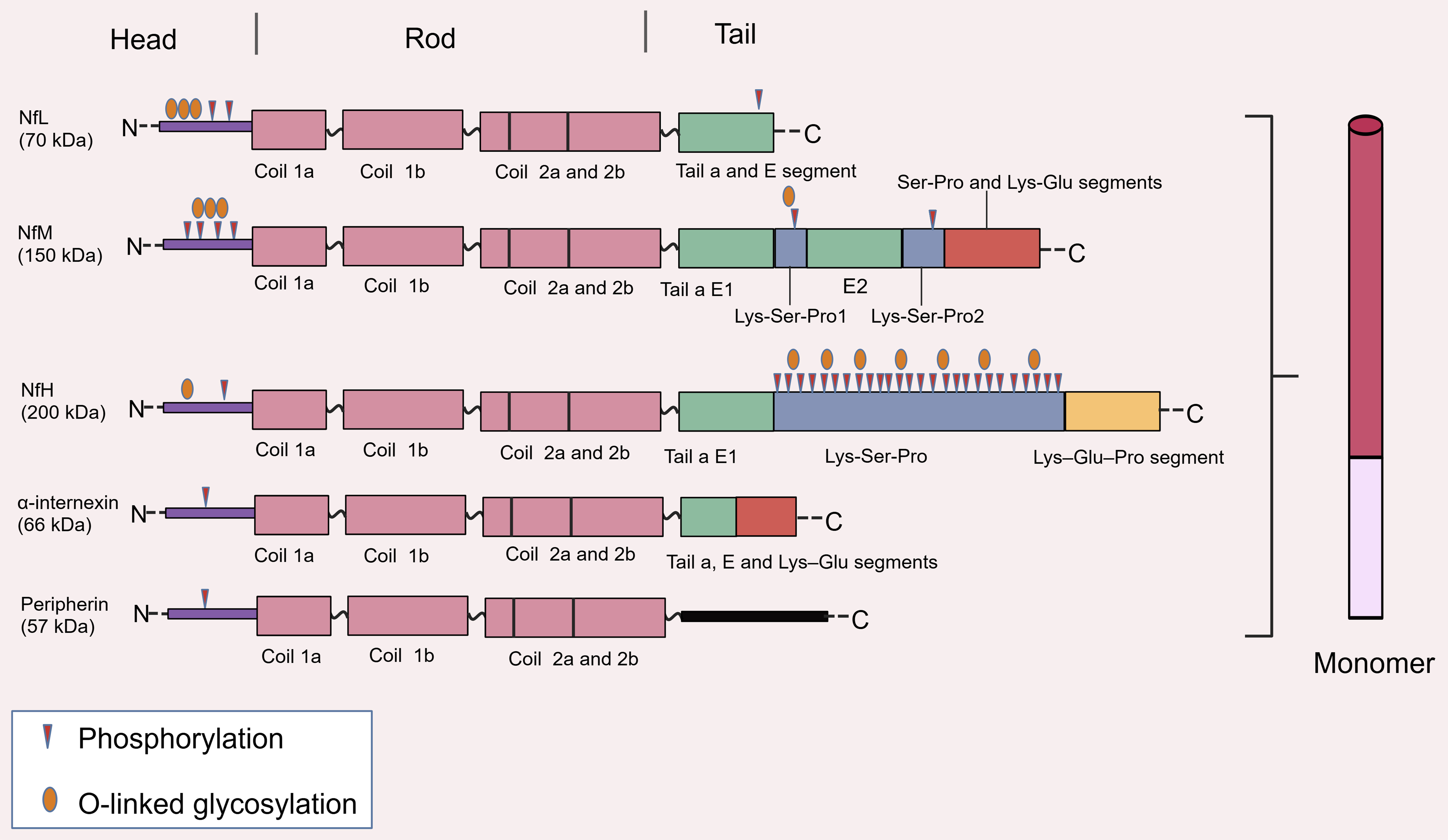
Figure 1. Types and structural organization of NF proteins. Adapted and modified from Yuan et al. (2017)[14], with additional graphical modifications and redesign by the authors. © Cold Spring Harbor Laboratory Press. Adapted with permission. Schematic illustration depicts the conserved three-domain architecture (N-terminal head, central rod, C-terminal tail) of NF subunits (NfL, NfM, NfH) and related neuronal intermediate filaments (α-internexin, peripherin), along with key post-translational modification sites including phosphorylation and O-linked glycosylation. Created with BioGDP.com. NF: Neurofilament; NfL: neurofilament light chain; NfM: neurofilament medium chain; NfH: neurofilament heavy chain.
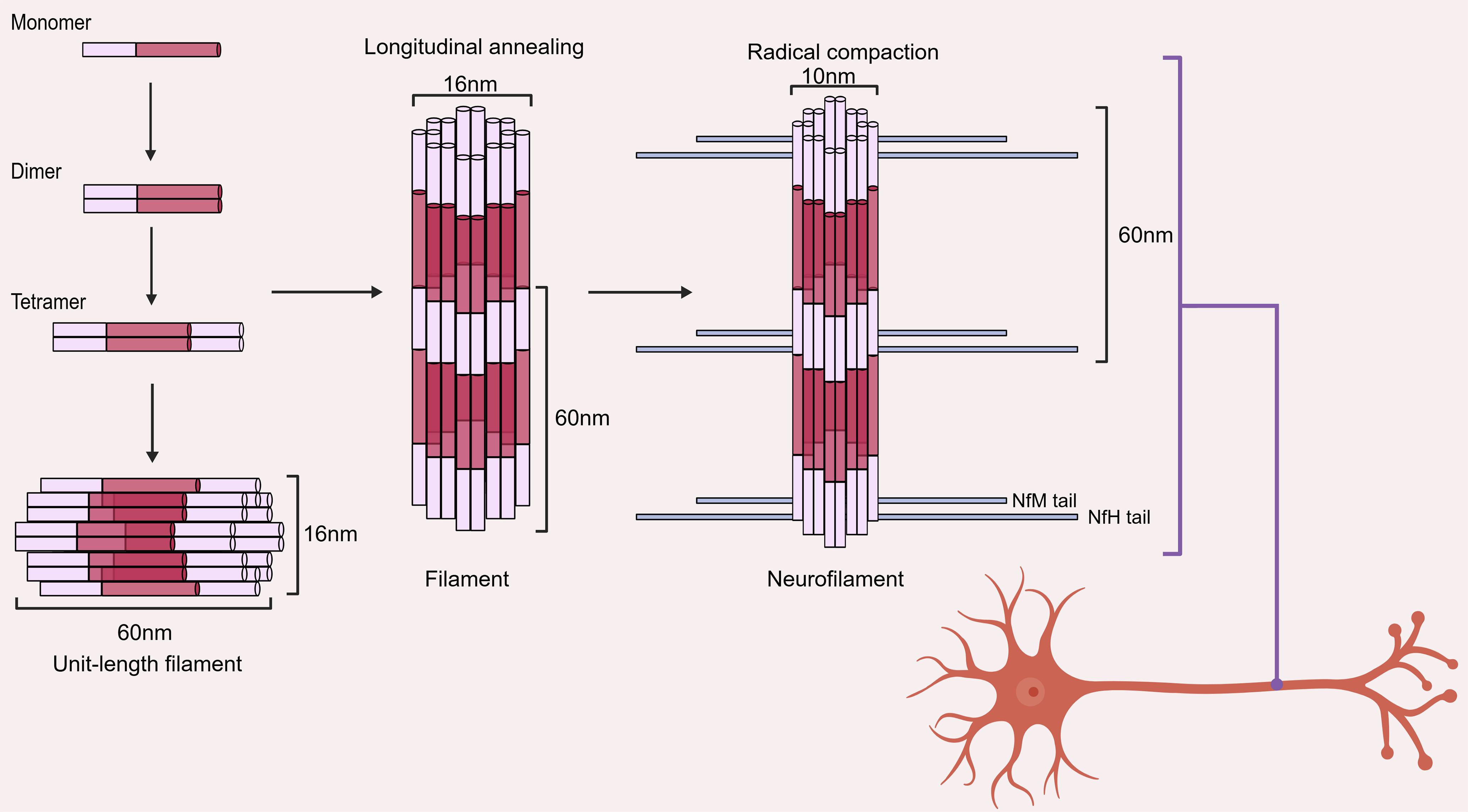
Figure 2. Hierarchical assembly process of NFs. Adapted and modified from Yuan et al. (2017)[14], with additional graphical modifications and redesign by the authors. © Cold Spring Harbor Laboratory Press. Adapted with permission. The diagram demonstrates the stepwise assembly pathway from monomeric subunits to mature NFs, including the formation of coiled-coil dimers, antiparallel tetramers, ULFs, and the final longitudinal annealing and radial compaction into 10 nm mature filaments. Created with BioGDP.com. NFs: Neurofilaments; ULFs: unit-length filaments; NfM: neurofilament medium chain; NfH: neurofilament heavy chain.
Under physiological conditions, NFs form the principal intermediate filament network within neurons, providing mechanical stability and maintaining axonal caliber and morphology[27]. By regulating axonal diameter, NFs directly influence nerve conduction velocity and ensure efficient signal transmission[28]. In addition, they interact with microtubules and motor proteins to coordinate axonal transport and intracellular organization, thereby contributing to organelle positioning and synaptic function, including the regulation of post-synaptic signaling and plasticity[29]. The stability of these structural and functional roles depends on tightly regulated molecular mechanisms. NFs assembly and spacing are modulated by post-translational modifications, particularly phosphorylation of the C-terminal domains of NfM and NfH, as well as glycosylation, ubiquitination, nitration, and oxidation[13]. These modifications regulate charge distribution, filament interaction, and cytoskeletal integration, thereby maintaining axonal integrity and transport dynamics[14]. Under pathological conditions, disruption of NFs homeostasis, such as abnormal phosphorylation or other post-translational alterations, can impair filament assembly and promote aggregation[30]. These changes interfere with axonal transport, compromise cytoskeletal stability, and ultimately contribute to axonal degeneration, a central feature of many neurodegenerative diseases[31]. As axonal damage progresses, NF proteins are released into the extracellular space and subsequently detected in CSF and blood[32]. This release provides the biological basis for their use as sensitive biomarkers of neuroaxonal injury, linking intracellular pathological processes to clinical monitoring of disease progression across a range of neurological and systemic conditions[33].
NFS AS BIOMARKERS IN NEURODEGENERATIVE DISEASES
In most neurodegenerative diseases, the initial triggering factors (such as abnormal Aβ and α-syn) act upstream. Once activated, these pathogenic processes initiate a cascade of pathological changes involving NFs[22]. Aberrant NFs can interact with pathological proteins to form co-aggregation complexes, not only exacerbating NF pathology, but also promoting the deposition of core pathogenic proteins, thereby emerging as a common pathological hallmark of multiple neurodegenerative disorders[25]. When NFs are disrupted, whether due to genetic defects, aberrant modifications, or co-aggregation with other proteins, they directly compromise their core functions, resulting in axonal instability, impaired transport, and disrupted signal conduction[13]. These pathological changes culminate in axonal degeneration and neuronal injury, a process that concurrently releases NFs into fluids (CSF and blood), establishing them as sensitive biomarkers reflecting the extent of neuroaxonal damage[34]. Therefore, NF pathology serves as a “key nexus” linking upstream pathology to downstream structural collapse.
NFs are both participants in the disease causal chain and non-specific damage biomarkers across diseases. Their elevated levels in body fluids are mainly a consequence of axonal damage, rather than the causative factor itself[16]. Clinically, the value of NFs may lie more in quantifying the extent and dynamic changes of neurodegeneration than in elucidating the underlying pathogenic mechanism.
DETECTION TECHNOLOGIES OF NEUROFILAMENT BIOMARKERS IN NEURODEGENERATIVE DISEASES
With the growing recognition of NFs as biomarkers of neurodegeneration, advances in detection technologies have fundamentally shifted their clinical utility from exploratory research tools to scalable blood-based indicators of neurodegenerative burden. Below we comprehensively summarize mainstream detection platforms, their performance characteristics, and key confounding factors that influence the reliability of measurement results.
Core detection platforms and sensitivity comparison
The evolution of NF detection technologies has followed a trajectory of progressively improved analytical sensitivity, enabling the transition from CSF-only measurement to reliable quantification in peripheral blood [Figure 3][16].
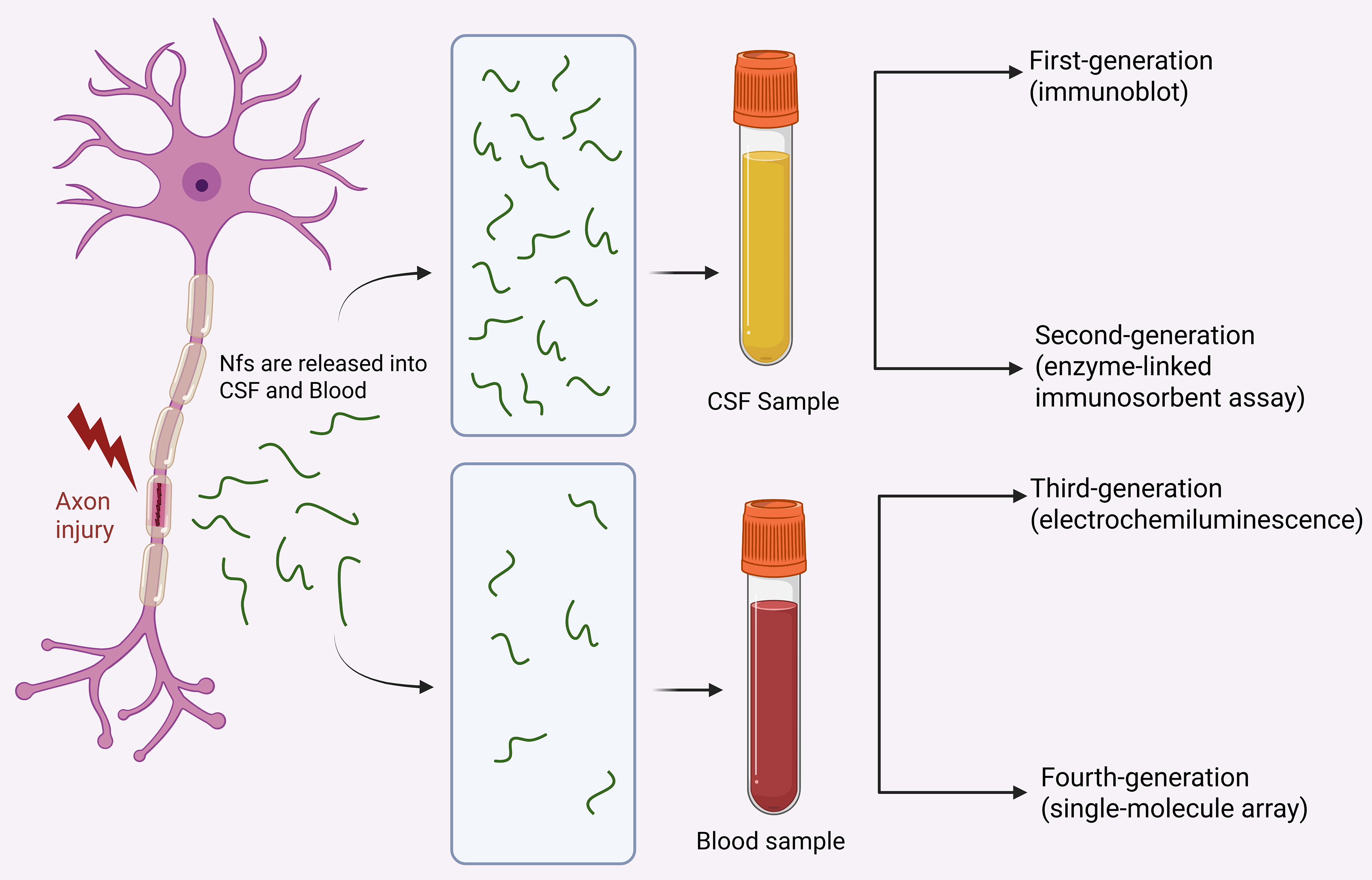
Figure 3. Release and detection of NFs after axonal injury. This figure was designed by the authors using BioRender. The schematic diagram shows that NF proteins are released into CSF and peripheral blood upon axonal damage in neurodegenerative conditions, and summarizes the evolutionary trajectory of detection technologies from early immunoblotting to ultrasensitive SiMoA assays. Created in BioRender. Zhao, X. (2026) https://BioRender.com/e86ov2c. NFs: Neurofilaments; CSF: cerebrospinal fluid; SiMoA: single-molecule array.
Enzyme-linked immunosorbent assay
As a classic second-generation immunoassay, enzyme-linked immunosorbent assay (ELISA) was the earliest widely adopted platform for NF quantification[35]. It relies on specific antibody-antigen binding coupled with colorimetric signal readout[36]. While ELISA performs adequately for measuring relatively high NF concentrations in CSF, its limited analytical sensitivity prevents accurate detection of low-abundance NFs in peripheral blood, restricting its application in minimally invasive clinical scenarios[37].
Electrochemiluminescence
As a third-generation detection platform, electrochemiluminescence (ECL) delivers substantial improvements in both analytical sensitivity and dynamic range compared with ELISA[38]. It relies on electrochemically triggered SULFO-TAG™ luminescent labels for signal generation, offering improved analytical precision and a wider linear detection range relative to conventional ELISA[39]. Despite this improved performance relative to ELISA, ECL still lacks sufficient analytical sensitivity to reliably quantify basal circulating NfL levels in healthy peripheral blood, with nearly half of healthy serum samples falling below its limit of detection in head-to-head validations[40].
SiMoA
The emergence of SiMoA marks a pivotal breakthrough in NF biomarker detection, representing the fourth generation of immunoassays with ultra-sensitive quantification capability at the single-molecule level[16,41]. With a limit of detection orders of magnitude lower than both conventional ELISA and ECL platforms, SiMoA enables reliable and reproducible measurement of low-abundance NfL in both serum and plasma[40]. This technological leap has resolved the long-standing sensitivity bottleneck of peripheral blood NF detection, directly accelerating the clinical translation of blood-based NF biomarkers and supporting the conduct of large-scale population cohorts and multicenter clinical trials worldwide[32].
Mass spectrometry
Mass spectrometry represents an emerging technology for NF detection, with unique advantages in proteoform differentiation and multidimensional molecular information output compared with antibody-based immunoassays[42]. It enables simultaneous measurement of multiple NF subunits (NfL, NfM, NfH) within a single analytical run[43]. It allows precise identification of post-translationally modified NF forms, such as phosphorylated NfH (pNfH), at the specific amino acid modification site level[44]. By covering multiple peptide regions across the full protein sequence, it can detect truncated proteolytic isoforms that cannot be recognized by single-epitope immunoassays, thus providing richer multidimensional biological information[45]. However, its widespread clinical application remains limited, and most mass spectrometry-based NF assays are still confined to research settings[42]. Most existing mass spectrometry assays for NFs involve multi-step pre-analytical workflows including immunoprecipitation, enzymatic digestion and chromatographic separation, resulting in relatively low sample throughput incompatible with high-volume routine clinical laboratories[36]. The vast majority of these methods are laboratory-developed tests without unified standardization protocols or commercialized kits, requiring specialized instrumentation and skilled technical personnel for implementation[46]. Mass spectrometry platforms incur higher instrument procurement, daily maintenance and per-sample testing costs compared with mature automated immunoassay platforms, which further hinders their large-scale clinical adoption from a health economics perspective[35].
Sensitivity comparison across platforms
Analytical sensitivity differs substantially across the platforms described above, which is a core source of inter-study heterogeneity[16]. SiMoA exhibits the highest sensitivity for blood NfL detection, followed by ECL, with conventional ELISA having the lowest sensitivity. The difference in detection sensitivity directly translates into differences in clinical applicability: ultra-sensitive platforms represented by SiMoA can stably detect NfL in serum of healthy people and patients with mild neuroaxonal injury, while conventional ELISA can hardly achieve reliable quantification of low-concentration blood NfL[40]. The fully automated chemiluminescence assays gradually applied in clinical routine have a moderate sensitivity level, which can meet the NfL quantification requirements of most neurological disease states while ensuring high-throughput and on-demand detection capacity[36,47]. As a candidate reference method to promote the standardization of NfL detection, liquid chromatography-tandem mass spectrometry currently has lower sensitivity than mainstream ultra-sensitive immunoassays, and is more suitable for CSF samples with higher endogenous NfL levels[46]. Systematic biases across different assay platforms can result in significant discrepancies in the absolute concentrations of NfL, highlighting the necessity of assay standardization and platform-specific reference ranges[36].
Factors influencing NF measurement results
Accurate interpretation of NF levels requires comprehensive consideration of multiple confounding factors, which can be categorized into physiological factors, comorbid conditions, and pre-analytical variability.
Physiological confounders
Age: Physiological factors are the critical determinant of NfL levels, which increase progressively with advancing age, particularly after 50 or 60 years, leading to marked differences in “normal ranges” across age groups[12].
Gender: In healthy populations, NfL levels in blood and CSF are generally higher in males than in females[19].
Body mass index (BMI): Higher BMI is often associated with lower NfL concentrations, likely due to a dilution effect caused by increased blood volume[32].
Renal dysfunction: Renal dysfunction, especially reduced estimated glomerular filtration rate, impairs the clearance of circulating NfL and results in non-specific elevations of blood NfL levels, representing an important confounder that must be accounted for[15].
Comorbidities
Comorbid conditions are another important factor that has to be considered. NfL is a non-specific marker of neuroaxonal injury and can be elevated in a wide range of neurological disorders, including AD, PD, stroke, and traumatic brain injury[48]. This substantially complicates the disease-specific interpretation of NfL[33]. In addition, certain systemic comorbidities-such as diabetes, cardiovascular disease, hypertension, and systemic inflammation-may indirectly alter NfL levels by affecting vascular health, neuroinflammation, or blood-brain barrier permeability[16].
Pre-analytical variability
The third factor that may affect detection results is the analytical and technical variability. Differences arising from detection platforms (e.g., SiMoA and ELISA), sample types (serum and plasma), and pre-analytical and analytical procedures can lead to variability of up to 50%[49]. Standardization of pre-analytical workflows is therefore a critical prerequisite for ensuring result comparability across studies and clinical centers.
To minimize these sources of variability, future efforts should prioritize the international harmonization of pre-analytical and analytical procedures, the development of standardized operating protocols, and cross-platform validation to improve the comparability and reproducibility of NfL measurements across laboratories. Furthermore, NfL results should be interpreted in conjunction with clinical assessment, neuroimaging findings, and complementary fluid biomarkers, such as glial fibrillary acidic protein (GFAP), rather than being used as a standalone biomarker. Such multidimensional approaches, together with individualized longitudinal monitoring when appropriate, may further enhance the clinical utility of NfL while reducing the influence of biological and technical confounding factors.
NF IMPAIRMENTS ACROSS NEURODEGENERATIVE DISEASES
NF proteins, as biomarkers for axonal damage, are found in a variety of neurological diseases. Below, we summarize the research progress of NF proteins in several neurodegenerative diseases.
AD
AD is a neurodegenerative disorder of the CNS marked by progressive cognitive decline and behavioral dysfunction[48]. The neuropathological hallmarks of AD are extracellular Aβ plaques and intracellular neurofibrillary tangles composed of hyperphosphorylated tau protein[50]. Importantly, neuropathological changes begin many years before the onset of overt clinical symptoms, and even after symptoms emerge[51], diagnosis may still be delayed by a year or more[52]. This temporal disconnect between pathology and clinical detection underscores a critical need for biomarkers to identify neurodegeneration in the preclinical phase.
Advances in proteomic technologies have facilitated the identification of candidate biomarkers for AD[53]. Longitudinal cohort studies confirm that blood NfL elevation occurs far in advance of clinical symptoms in autosomal dominant AD. In the DIAN cohort, the longitudinal change rate of serum NfL (sNfL) can discriminate mutation carriers approximately 16 years before estimated symptom onset[54]. In the Colombian PSEN1 E280A mutation kindred, plasma NfL divergence between carriers and non-carriers is detectable as early as 22 years before mild cognitive impairment onset[55]. These findings indicate that NFs alterations reflect early neurodegenerative processes rather than late-stage consequences.
Basic research indicates that calpain, a key protease involved in NF metabolism, can trigger the disintegration of the NF network upon activation, a mechanism that may contribute to axonal degeneration in AD[14]. Meanwhile, proline-directed kinases that catalyze tau protein hyperphosphorylation can also phosphorylate the tail domains of NfM and NfH, suggesting that in AD, tau pathology and neurofilamentopathy may be driven by common upstream kinases, forming a mutually exacerbating vicious cycle[56].
Interpretation of NfL levels in AD must account for their lack of disease specificity[42]. Although NfL concentrations increase with normal aging, patients with neurodegenerative diseases, including AD, exhibit higher absolute levels or faster rates of increase compared with age-matched healthy individuals[57]. Nonetheless, the ability of NfL to distinguish AD from other dementias remains limited. This is largely because NfL is elevated across multiple neurological conditions, including multiple sclerosis and frontotemporal dementia (FTD)[58]. Consequently, while NfL reflects neuronal injury, its diagnostic utility as a standalone marker for AD is modest.
Despite these limitations, NfL may be more valuable as a marker of disease severity and progression rather than diagnosis[16]. Elevated NfL levels in CSF and blood correlate with poorer cognitive performance, greater neurodegeneration, and accelerated disease progression[32]. Cross-sectional studies have demonstrated associations between higher plasma NfL levels and more severe medial temporal lobe atrophy and reduced metabolic activity in AD-related cortical regions[7]. Longitudinal analyses further show that elevated baseline NfL predicts faster hippocampal and temporal cortical atrophy and greater declines in fluorodeoxyglucose uptake over 15-30 months[57]. While NfL alone cannot establish an AD diagnosis, its prognostic and monitoring value is significantly enhanced when combined with established plasma biomarkers such as Aβ42, Aβ40, p-tau217, and p-tau181[56].
Other NF subunits may also contribute complementary biochemical and diagnostic information. A multi-cohort CSF proteomic study found that CSF NfM (cNfM) levels are significantly elevated in AD patients compared with cognitively healthy controls, with a trend of increase observed in the MCI stage. As an exploratory proteomic finding, its capacity to differentiate AD from MCI and its disease specificity require further validation in larger independent cohorts[48]. pNfH, while well-established in motor neuron disease diagnostics, may also reflect neurodegenerative processes in dementia. In a memory clinic cohort of 188 patients, CSF pNfH and NfL (cNfL) levels were significantly elevated across diagnostic groups compared with controls, and plasma pNfH was also increased[58].
Taken together, these findings suggest that in AD, NFs function less as disease-specific diagnostic markers and more as indicators of neurodegenerative intensity and progression. Their greatest clinical value may therefore lie in monitoring disease dynamics and in complementing core AD biomarkers within multimodal diagnostic frameworks.
PD
PD is a common neurodegenerative disorder that predominantly affects middle-aged and elderly individuals. Its clinical features include resting tremor, bradykinesia, muscle rigidity, and postural and gait disturbances[59]. Atypical parkinsonian syndromes (APS) are characterized by rapid disease progression, the presence of parkinsonian features accompanied by other neurological manifestations, and a poor response to conventional anti-PD medications[33]. There is significant overlap in the clinical presentations of PD and APS, especially in the early stages of the disease, highlighting the urgent need for objective biomarkers that can differentiate these diseases based on the extent of neurodegeneration rather than clinical manifestations alone[16].
Numerous cross-sectional and longitudinal studies have consistently shown that CSF and blood NfL levels are significantly higher in APS patients than in PD patients, with elevated levels observed across all APS subgroups[60,61]. Receiver operating characteristic analysis revealed that the areas under the curve (AUCs) for distinguishing PD from APS using CSF and blood NfL levels reached 0.94 and 0.87, respectively[60]. A possible mechanistic explanation is that APS is associated with faster and more extensive neurodegeneration, with more severe involvement of large-diameter myelinated axons in the subcortical brain compared to PD, and the degeneration of myelinated axons may release more NfL than neuronal soma degeneration[59]. Furthermore, combining clinical features, neuroimaging, and biomarkers, including NfL and other more disease-specific markers such as α-syn seeding activity, may be more effective than any single modality[61].
Currently, research on NfL as a biomarker for neurodegeneration in PD is still ongoing[16]. Existing evidence suggests that elevated cNfL levels in certain PD subgroups may reflect a more aggressive and extensive neurodegenerative process[60]. In the population-based New Parkinsonism in Umeå cohort, higher cNfL levels were associated with more severe PD symptoms assessed by clinical scales (e.g., Hoehn and Yahr stage and UPDRS scores) and shorter survival[59]. In two longitudinal biomarker cohorts for PD progression, sNfL levels were elevated in PD patients compared to healthy controls and increased over time and with age[33]. Additionally, in a cohort including PD and Parkinson’s disease dementia, both plasma NfL and cNfL levels were significantly correlated with motor dysfunction, Mini-Mental State Examination scores, and orthostatic hypotension[61]. The relationship between NfH levels and PD clinical progression remains unclear; a post-hoc cohort study demonstrated that elevated baseline CSF NfH levels in early PD patients predicted faster motor and cognitive decline, and the association between NfH and cognitive decline was stronger than that observed for NfL[62].
Although NfL lacks disease specificity for PD, it represents one of the most promising blood-based biomarker candidates for the disease. Further research is needed to determine whether CSF or blood NfL can be used for longitudinal monitoring of PD progression and whether combining age, more disease-specific biomarkers, and simplified clinical subtyping scoring systems can yield clinically useful prognostic tools[63].
DLB
DLB is one of the major causes of dementia, with an incidence second only to AD and FTD[16]. Unlike AD, there are currently no blood biomarkers that can directly reflect the underlying pathological changes in DLB, which poses a significant diagnostic challenge, especially in the early stages of the disease, when clinical symptoms overlap with other dementia syndromes[64].
NfL is elevated in the CSF and plasma of DLB patients; however, its levels are generally comparable to those in AD patients and lower than in FTD patients, limiting its utility as a standalone tool for differential diagnosis[65]. NfL levels are not merely passive indicators of axonal damage but also molecular signatures resulting from the combined effects of α-syn pathology, axonal transport disorders, and genetic background[14]. Aberrant activation of calpain serves as a critical driver of neuronal injury across multiple neurodegenerative diseases. In DLB, calpain mediates site-specific cleavage of α-syn aggregation. The resulting truncated isoforms display enhanced aggregation propensity and form the core component of Lewy bodies, accelerating protein misfolding and aggregation propagation and driving dopaminergic neuron degeneration. This pathological alteration has been validated in human brain tissue specimens[66]. Calpain also mediates broad-spectrum axonal injury and degeneration via the degradation of axonal cytoskeletal proteins: NfL, NfM and NfH, act as calpain substrates, with NfM being the most susceptible to proteolytic cleavage and undergoing degradation as early as the initial phase of calcium overload. Studies in multiple animal models of neural injury and neurodegeneration demonstrate that calpain-mediated NF hydrolysis disrupts axonal cytoskeletal integrity, subsequently triggering axonal transport dysfunction and axonal degeneration, and represents a shared downstream pathway of neuronal damage[67].
Evidence from the Amsterdam Dementia Cohort shows that combined detection of plasma p-tau181, NfL, and GFAP can improve the differential diagnosis between AD and non-AD dementias, including FTD and DLB, with AUCs of 0.8[64]. While NFs alone cannot resolve the diagnostic complexity of DLB, they may still play a supportive role in stratifying neurodegeneration intensity and improving differential diagnosis within emerging precision neurology frameworks.
FTD
FTD encompasses multiple clinical syndromes, including behavioral variant FTD, nonfluent variant primary progressive aphasia, and semantic variant primary progressive aphasia, each of which differs in its dominant cognitive and behavioral features[68]. This clinical heterogeneity reflects underlying pathological and genetic diversity, with common causative mutations involving chromosome 9 open reading frame 72 (C9orf72), progranulin (PGRN), and microtubule-associated protein tau (MAPT)[16].
This heterogeneity poses challenges for disease stratification and monitoring. As a core structural element of the neuronal axonal cytoskeleton, changes in NfL levels not only passively reflect neuronal damage but are also closely related to pathological mechanisms driven by different gene mutations[14]. Animal models and studies of molecular mechanisms provide key evidence for this. In C9orf72-associated FTD, toxic dipeptide repeat proteins derived from hexanucleotide repeat expansions disrupt cytoskeletal homeostasis and impair axonal transport by altering microtubule-associated protein regulation and compromising NF network integrity[13,69]. In PGRN mutations, haploinsufficiency of PGRN triggers lysosomal dysfunction and TDP-43 pathology, and TDP-43, as a binding protein for NfL mRNA, can directly affect the synthesis and homeostasis of NF proteins through its abnormal aggregation[70]. In MAPT mutations, hyperphosphorylated tau protein detaches from microtubules, secondarily disrupting the integrity of the NFs network[14]. These gene-specific mechanisms provide a profound explanation for the NfL kinetic characteristics observed clinically.
In one FTD-focused study, cNfL concentrations in patients were more than threefold higher than in cognitively unimpaired controls, with an increase greater than that observed in AD[64]. This pronounced elevation may reflect the more extensive frontotemporal degeneration and subcortical involvement characteristic of FTD[17]. A strong correlation has been observed between CSF and blood NfL levels, and similar trends have been reported in plasma, where FTD patients exhibited significantly higher plasma NfL levels than all other diagnostic groups [vs. progressive supranuclear palsy (PSP), DLB, AD, and suspected non-AD physiopathology (SNAP), and corticobasal syndrome (CBS); all P < 0.001][65]. Blood NfL levels in FTD patients vary depending on the underlying genetic mutations, which directly reflects the heterogeneity at the mechanistic level mentioned above[71]. The highest levels are observed in PGRN mutation carriers, reflecting intense TDP-43-driven axonal disintegration, whereas the lowest levels are observed in MAPT mutation carriers, consistent with axonal damage secondary to Tau pathology[72].
The magnitude of presymptomatic NfL elevation has important prognostic implications[16]. In the GENFI and ALLFTD cohort studies, the rate of change in sNfL peaked during the prodromal phase as individuals transitioned from asymptomatic to symptomatic disease[70,73]. Higher sNfL concentrations were consistently associated with greater disease severity and poorer performance across multiple domains, including global cognition, social behavior, language, and executive function[71].
Nevertheless, diseases within the FTD spectrum remain highly heterogeneous, and differences between familial and sporadic FTD further complicate interpretation[68]. Consequently, it has been proposed that NfL be used in combination with other pathophysiological biomarkers to more reliably reflect disease progression[70]. Such multimodal approaches may improve the prediction of disease trajectories and enhance the evaluation of disease-modifying therapies in clinical trials.
Huntington’s disease
Huntington’s disease (HD) is a progressive autosomal dominant inherited neurodegenerative disorder characterized by motor dysfunction, psychiatric abnormalities, and cognitive impairment. The disease is caused by a CAG trinucleotide repeat expansion within the huntingtin (HTT) gene, leading to the production of mutant huntingtin protein (mHTT)[74]. Although various CSF biomarkers have been proposed for HD, only a few, such as mHTT, have shown clear associations with clinical phenotypes[75].
In R6/2 mouse models expressing human mHTT, researchers observed significant increases in NfL levels in CSF and blood as neuronal damage occurred, with the magnitude of the increase directly correlating with the extent of axonal degeneration[76]. An independent CSF validation cohort from London further demonstrated a high correlation between CSF and plasma NfL concentrations, indicating that blood measurements can serve as a reliable window into CNS damage[77].
Multiple case-control and longitudinal studies have shown that cNfL levels are elevated in HD patients and correlate with clinical severity[78]. However, compared to cNfL and mHTT, plasma NfL shows the strongest association with clinical disease severity[77]. Baseline plasma NfL levels have been demonstrated to predict multiple aspects of subsequent disease progression, including regional and global brain atrophy rates, particularly in disease-related brain regions[79]. Cognitive decline, assessed by measures such as the Unified Huntington’s Disease Rating Scale and the Symbol Digit Modalities Test, is associated with higher plasma NfL levels and poorer cognitive performance, as well as smaller brain volumes[75]. Furthermore, by analyzing the temporal changes of various CSF markers, research found that the elevation of NfL precedes that of neuroinflammatory markers; this key finding supports the pathological model of HD: primary axonal damage (reflected by NfL) caused by the direct toxicity of mHTT is the initiating factor of the disease, while subsequent glial cell activation and neuroinflammatory responses are likely secondary responses to the initial neuronal damage[80]. Longitudinal analysis indicates that in premanifest mutation carriers, baseline plasma NfL can independently predict the risk of disease conversion within three years, as well as future cognitive decline, multi-site brain atrophy, and ventricular enlargement[75].
In clinical research settings, employing NfL as an outcome measure of treatment efficacy may significantly improve statistical power, potentially outperforming the use of CSF mHTT alone[77]. In future clinical trials of disease-modifying therapies, a reduction in NfL levels post-intervention would directly indicate a protective effect on neuronal axons and serve as a sensitive surrogate marker for efficacy evaluation[78]. There is still a lack of large, well-phenotyped cohorts that include both plasma and CSF samples. Therefore, long-term longitudinal data are needed to characterize the trajectory of these biomarkers throughout the disease process and to directly compare their value in predicting disease progression.
Amyotrophic lateral sclerosis
Amyotrophic lateral sclerosis (ALS) is a fatal neurodegenerative disease characterized by the progressive degeneration of both upper and lower motor neurons[81]. Average survival is only two to five years following diagnosis. Currently, early diagnosis remains limited, highlighting a critical unmet need for objective biomarkers that detect disease activity earlier[82]. In this context, NfL has emerged as a promising candidate biomarker. Elevated NfL levels have even been proposed as susceptibility biomarkers, with increased concentrations observed in asymptomatic carriers of pathogenic ALS-associated mutations potentially reflecting an increased risk of disease onset[28].
Proteomic analysis of autopsy spinal cord tissues from 8 patients found that although NF proteins content was significantly reduced in degenerated spinal cord tissues, their levels in CSF were significantly elevated. This phenomenon suggests that the elevation of NFs in CSF may primarily originate from the release of damaged neurons[16]. Although NFs are not disease-specific biomarkers, meta-analyses of evidence indicate that their concentrations in both CSF and blood are generally higher in patients with ALS compared to other neurodegenerative diseases[83]. This phenomenon may be explained by the selective vulnerability and rapid degeneration of large-caliber motor neurons, resulting in a substantial release of NF proteins into CSF and peripheral blood[15].
NFs levels provide valuable prognostic insight into disease progression. Elevated NfL and NfH concentrations correlate with a more rapid decline in ALS Functional Rating Scale-Revised scores and reduced survival[84]. Predictive modeling analyses by Witzel’s team have demonstrated that higher NFs levels are consistent predictors of accelerated disease progression and shorter life expectancy[15]. While the precise onset of NFs elevation remains under investigation, current evidence suggests that NfL levels increase during early symptomatic stages before plateauing as the disease advances[28].
To summarize, NFs elevation indicates neuroaxonal injury; it lacks disease specificity; therefore, it cannot independently establish a diagnosis of ALS[85]. Longitudinal trajectories of NFs levels remain inconsistent across studies, potentially reflecting heterogeneity in disease onset site, progression rate, or genetic background[11]. Despite these challenges, NFs have emerged as the most robust prognostic biomarkers currently available for ALS[86]. The antisense oligonucleotide tofersen reduces SOD1 protein levels in the CNS, slows disease progression, and significantly decreases sNfL levels in SOD1 mouse models. In phase 3 clinical trials of patients with SOD1-mutated ALS, tofersen treatment also results in robust and sustained reductions in plasma NfL, supporting its value as a pharmacodynamic biomarker for therapeutic efficacy[82,87]. These preclinical and clinical findings collectively establish NfL not only as a sensitive biomarker of axonal damage, but also as a pharmacodynamic indicator for verifying target engagement and therapeutic efficacy in ALS drug development. Future efforts aimed at assay harmonization, reference standardization, and integration with clinical parameters will further enhance their clinical utility[88].
CONCLUSION AND FUTURE PERSPECTIVES
NFs, particularly NfL, have been extensively studied as sensitive biomarkers of neuroaxonal injury across a wide range of neurological disorders. With the advent of high-sensitivity detection technologies, such as SiMoA assays, reliable measurement of NfL in blood has become feasible, highlighting its considerable potential for disease screening, longitudinal monitoring, and assessment of treatment response. NfL levels are significantly elevated in multiple neurodegenerative diseases, including AD, PD, DLB, and are closely associated with disease progression, the extent of neurodegeneration, and clinical prognosis [Table 1].
NF detection in neurodegenerative diseases
| Disease | Pathogenesis involvement | Biomarker findings | Diagnostic value |
| AD | Tau kinases phosphorylate NfM, NfH[56]; calpain-mediated NFs degradation[14]; interactions with amyloid-β and tau pathology contribute to axonal degeneration[56] | Blood and CSF NfL are elevated years before symptom onset[16]; cNfM is elevated in AD compared with controls, with preliminary discriminatory potential[48] | Limited diagnostic specificity, requires combination with Aβ, p-tau[42,56]; predicts hippocampal atrophy and rate of cognitive decline[7,57] |
| PD | Axonal degeneration of dopaminergic neurons, more pronounced axonal injury in APS[59] | CSF and blood NfL are higher in APS than in PD[60,61]; CSF NfH predicts faster motor and cognitive decline in early PD[62] | Differentiation of PD and APS (AUCs up to 0.94)[60]; higher baseline levels predict faster motor and cognitive deterioration[59,62] |
| DLB | Calpain‑mediated site‑specific cleavage of α‑syn promotes its aggregation and Lewy body formation[66]; calpain‑dependent degradation of NF subunits induces NFs fragmentation and axonal transport dysfunction[67] | Elevated plasma and CSF NfL levels overlap with AD but are generally lower than FTD[16,65] | Limited differential diagnostic value, a combination of p-tau181, NfL, and GFAP improves diagnostic accuracy[64] |
| FTD | C9orf72 repeat expansions disrupt NF network integrity and impair axonal transport via toxic dipeptide repeat proteins[13,69]; PGRN mutations affect NfL synthesis and homeostasis[70,72]; MAPT mutations disrupt NFs network integrity[14] | Markedly elevated plasma and CSF NfL, substantially greater than in AD[64,65]; the highest levels observed in PGRN mutation carriers[71,72] | Disease severity marker, reflects the severity of frontotemporal atrophy[16]; predicts phenoconversion and disease progression[70,71] |
| HD | mHTT toxicity induces axonal degeneration and NFs release, which precedes the occurrence of neuroinflammation[76,80] | Plasma NfL correlates with clinical severity more strongly than CSF NfL or mHTT[77] | Predicts 3-year risk of disease onset[75], and rate of brain atrophy[79]; useful in clinical trial monitoring[75,77] |
| ALS | Rapid degeneration of large motor neurons leads to massive NFs release[15] | Highest NfL levels among neurodegenerative diseases[83]; spinal cord NFs content decreases while fluid levels surge[16] | Strong prognostic biomarker[86]; predicts survival[84], and treatment response[82] |
In contrast, research on NfM and NfH remains relatively limited. Differences in molecular weight, phosphorylation status, assay availability, and the scarcity of large-scale datasets have constrained their investigation, resulting in a predominant focus on NfL[15]. Future studies should prioritize developing more sensitive detection methods and conducting systematic investigations of NfM and NfH to comprehensively elucidate their pathological significance and clinical utility.
Despite its promise, several challenges hinder the clinical implementation of NfL. As a non-specific marker of neuroaxonal damage, NfL is elevated across a broad spectrum of neurological conditions, limiting its utility for single-disease differential diagnosis. In addition, baseline NfL levels are substantially influenced by confounding factors such as age, renal function, and BMI. Recent studies have demonstrated that plasma NfL concentrations increase progressively with physiological aging, highlighting the necessity of age-stratified reference intervals for accurate clinical interpretation. The establishment of reference values based on healthy populations therefore represents an important step toward the routine clinical application of blood NfL. Methodological variability across analytical platforms and differences between sample types further compromise data comparability, impeding the establishment of standardized reference ranges. Furthermore, recent international investigations have revealed substantial heterogeneity among expert centers regarding assay platforms, interpretation strategies, reference values, and laboratory reporting, underscoring the urgent need for international harmonization before NfL can be fully integrated into routine neurological practice. Consequently, interpretation of NfL levels must be contextualized within the clinical setting and adjusted for individual-specific factors.
Looking forward, advancing assay standardization and establishing cross-platform harmonization frameworks represent critical priorities. Beyond improving analytical performance, future efforts should focus on developing internationally harmonized reference intervals, standardized interpretation strategies, and unified laboratory reporting systems to facilitate the routine clinical implementation of NfL across different healthcare settings. From a clinical perspective, further validation of NfL as a reliable endpoint in clinical trials and as a prognostic biomarker is required. Integrating NfL with neuroimaging modalities and other fluid biomarkers may enable the development of individualized disease prediction models. Through interdisciplinary collaboration, international standardization initiatives, and large-scale longitudinal studies, NF proteins have the potential to evolve from promising research biomarkers into indispensable tools for precision diagnosis, therapeutic monitoring, and personalized management of neurodegenerative diseases.
DECLARATIONS
Acknowledgments
The Graphical Abstract was created with BioRender. Zhao, X. (2026) https://BioRender.com/ztvm99a.
Authors’ contributions
Conducted the literature retrieval and data sorting, drafted the manuscript, and completed the design and production of all figures and tables: Zhao X
Clarified the overall academic logic and conceptual framework, provided critical intellectual suggestions for content revision, and polished the academic writing and grammar of the manuscript: Hu Y
Both authors have read and approved the final version of the manuscript for publication.
Availability of data and materials
Not applicable.
AI and AI-assisted tools statement
Not applicable.
Financial support and sponsorship
This work was supported by the National Natural Science Foundation of China (Grant No. 82501716).
Conflicts of interest
Both authors declared that there are no conflicts of interest.
Ethical approval and consent to participate
Not applicable.
Consent for publication
Not applicable.
Copyright
© The Author(s) 2026.
REFERENCES
1. Wang JT, Xu G, Ren RJ, et al. The impacts of health insurance and resource on the burden of Alzheimer’s disease and related dementias in the world population. Alzheimers Dement. 2023;19:967-79.
3. Jia YJ, Ge YJ, Li B, et al. Advances in Alzheimer’s disease: mechanistic insights and therapeutic targets. Sci China Life Sci. 2026;69:2543-74.
4. Hatano T, Okuzumi A, Matsumoto G, Tsunemi T, Hattori N. α-Synuclein: a promising biomarker for Parkinson’s disease and related disorders. J Mov Disord. 2024;17:127-37.
5. Atri A, Dickerson BC, Clevenger C, et al. The Alzheimer’s Association clinical practice guideline for the diagnostic evaluation, testing, counseling, and disclosure of suspected Alzheimer’s disease and related disorders (DETeCD-ADRD): Validated clinical assessment instruments. Alzheimers Dement. 2025;21:e14335.
6. Xu J, Xu X, Guo X, et al. Improving reliability of movement assessment in Parkinson’s disease using computer vision-based automated severity estimation. J Parkinsons Dis. 2025;15:349-60.
7. Gallingani C, Carbone C, Tondelli M, Zamboni G. Neurofilaments light chain in neurodegenerative dementias: a review of imaging correlates. Brain Sci. 2024;14:272.
8. Zetterberg H, Blennow K. Moving fluid biomarkers for Alzheimer’s disease from research tools to routine clinical diagnostics. Mol Neurodegener. 2021;16:10.
9. Rissardo JP, Fornari Caprara AL. Alpha-synuclein seed amplification assays in Parkinson’s disease: a systematic review and network meta-analysis. Clin Pract. 2025;15:107.
10. Kobayashi N. Editorial: Blood, cerebrospinal fluid, and vascular biomarkers for dementia. Front Dement. 2026;5:1747825.
11. Turner MR, Thompson AG, Teunissen CE. Blood level of neurofilament light chain as a biomarker for neurological disorders. BMJ Med. 2025;4:e000958.
12. Bar-Or A, Nicholas J, Feng J, Sorrell F, Cascione M. Exploring the clinical utility of neurofilament light chain assays in multiple sclerosis management. Neurol Neuroimmunol Neuroinflamm. 2025;12:e200427.
13. Devarakonda SS, Basha S, Pithakumar A, et al. Molecular mechanisms of neurofilament alterations and its application in assessing neurodegenerative disorders. Ageing Res Rev. 2024;102:102566.
14. Yuan A, Rao MV, Veeranna, Nixon RA. Neurofilaments and neurofilament proteins in health and disease. Cold Spring Harb Perspect Biol. 2017;9:a018309.
15. Witzel S, Mayer K, Oeckl P. Biomarkers for amyotrophic lateral sclerosis. Curr Opin Neurol. 2022;35:699-704.
16. Khalil M, Teunissen CE, Lehmann S, et al. Neurofilaments as biomarkers in neurological disorders - towards clinical application. Nat Rev Neurol. 2024;20:269-87.
17. Khalil M, Teunissen CE, Otto M, et al. Neurofilaments as biomarkers in neurological disorders. Nat Rev Neurol. 2018;14:577-89.
18. Götze K, Vrillon A, Dumurgier J, et al. Plasma neurofilament light chain as prognostic marker of cognitive decline in neurodegenerative diseases, a clinical setting study. Alzheimers Res Ther. 2024;16:231.
19. Kölliker Frers RA, Otero-Losada M, Kobiec T, et al. Multidimensional overview of neurofilament light chain contribution to comprehensively understanding multiple sclerosis. Front Immunol. 2022;13:912005.
20. Simrén J, Andreasson U, Gobom J, et al. Establishment of reference values for plasma neurofilament light based on healthy individuals aged 5-90 years. Brain Commun. 2022;4:fcac174.
21. Delaby C, Ladang A, Martinez-Yriarte J, et al. Clinical use and reporting of neurofilament quantification in neurological disorders: a global overview. Alzheimers Dement. 2025;21:e70343.
22. van Asperen JV, Kotaich F, Caillol D, Bomont P. Neurofilaments: novel findings and future challenges. Curr Opin Cell Biol. 2024;87:102326.
23. Virata MCA, Catahay JA, Lippi G, Henry BM. Neurofilament light chain: a biomarker at the crossroads of clarity and confusion for gene-directed therapies. Neurodegener Dis Manag. 2024;14:227-39.
24. Kotaich F, Caillol D, Bomont P. Neurofilaments in health and Charcot-Marie-Tooth disease. Front Cell Dev Biol. 2023;11:1275155.
25. Bomont P. The dazzling rise of neurofilaments: physiological functions and roles as biomarkers. Curr Opin Cell Biol. 2021;68:181-91.
26. Uchida A, Peng J, Brown A. Regulation of neurofilament length and transport by a dynamic cycle of phospho-dependent polymer severing and annealing. Mol Biol Cell. 2023;34:ar68.
27. Ding EA, Kumar S. Neurofilament biophysics: from structure to biomechanics. Mol Biol Cell. 2024;35:re1.
28. Sanchez-Tejerina D, Llaurado A, Sotoca J, et al. Biofluid biomarkers in the prognosis of amyotrophic lateral sclerosis: recent developments and therapeutic applications. Cells. 2023;12:1180.
29. Zhu PP, Hung HF, Batchenkova N, et al. Transverse endoplasmic reticulum expansion in hereditary spastic paraplegia corticospinal axons. Hum Mol Genet. 2022;31:2779-95.
30. De Paoli LF, Kirkcaldie MTK, King AE, Collins JM. Neurofilament heavy phosphorylated epitopes as biomarkers in ageing and neurodegenerative disease. J Neurochem. 2025;169:e16261.
31. Bittner S, Oh J, Havrdová EK, Tintoré M, Zipp F. The potential of serum neurofilament as biomarker for multiple sclerosis. Brain. 2021;144:2954-63.
32. Teunissen CE, Verberk IMW, Thijssen EH, et al. Blood-based biomarkers for Alzheimer’s disease: towards clinical implementation. Lancet Neurol. 2022;21:66-77.
33. Mollenhauer B, Dakna M, Kruse N, et al. Validation of serum neurofilament light chain as a biomarker of Parkinson’s disease progression. Mov Disord. 2020;35:1999-2008.
34. Sharma P, Giri A, Tripathi PN. Emerging trends: neurofilament biomarkers in precision neurology. Neurochem Res. 2024;49:3208-25.
35. Bavato F, Barro C, Schnider LK, et al. Introducing neurofilament light chain measure in psychiatry: current evidence, opportunities, and pitfalls. Mol Psychiatry. 2024;29:2543-59.
36. La Civita E, Nicolella V, Fiorenza M, et al. Advancing clinical use of neurofilament light chain: translational insights from research to routine practice. Biomark Insights. 2025;20:11772719251364018.
37. Alagaratnam J, von Widekind S, De Francesco D, et al. Correlation between CSF and blood neurofilament light chain protein: a systematic review and meta-analysis. BMJ Neurol Open. 2021;3:e000143.
38. Alirezaei Z, Pourhanifeh MH, Borran S, Nejati M, Mirzaei H, Hamblin MR. Neurofilament light chain as a biomarker, and correlation with magnetic resonance imaging in diagnosis of CNS-related disorders. Mol Neurobiol. 2020;57:469-91.
39. Ulndreaj A, Sohaei D, Thebault S, et al. Quantitation of neurofilament light chain protein in serum and cerebrospinal fluid from patients with multiple sclerosis using the MSD R-PLEX NfL assay. Diagnosis. 2023;10:275-80.
40. Kuhle J, Barro C, Andreasson U, et al. Comparison of three analytical platforms for quantification of the neurofilament light chain in blood samples: ELISA, electrochemiluminescence immunoassay and Simoa. Clin Chem Lab Med. 2016;54:1655-61.
41. Barro C, Chitnis T, Weiner HL. Blood neurofilament light: a critical review of its application to neurologic disease. Ann Clin Transl Neurol. 2020;7:2508-23.
42. Daponte A, Koros C, Skarlis C, et al. Neurofilament biomarkers in neurology: from neuroinflammation to neurodegeneration, bridging established and novel analytical advances with clinical practice. Int J Mol Sci. 2025;26:9739.
43. Petzold A. Proteolysis-based biomarker repertoire of the neurofilament proteome. J Neurochem. 2025;169:e70023.
44. Rudrabhatla P, Jaffe H, Pant HC. Direct evidence of phosphorylated neuronal intermediate filament proteins in neurofibrillary tangles (NFTs): phosphoproteomics of Alzheimer’s NFTs. FASEB J. 2011;25:3896-905.
45. Coulton JB, He Y, Barthélemy NR, Jiang H, Holtzman DM, Bateman RJ. Multi-peptide characterization of plasma neurofilament light chain in preclinical and mild Alzheimer’s disease. Brain Commun. 2024;6:fcae247.
46. Coppens S, Vialaret J, Mondésert E, et al. Development of a novel liquid chromatography coupled to multiple reaction monitoring (LC-MRM) assay for the quantification of neurofilament light chain in cerebrospinal fluid and comparison with ultra-sensitive immunoassay: a step toward standardization. Clin Chem. 2026;72:478-87.
47. Mondésert E, Schraen-Maschke S, Quadrio I, et al. A French multicenter analytical evaluation of the automated Lumipulse G sNfL blood assay (Fujirebio®) and its comparison to four other immunoassays for serum neurofilament light chain assessment in clinical settings. Clin Chim Acta. 2025;565:120007.
48. Bergström S, Remnestål J, Yousef J, et al. Multi-cohort profiling reveals elevated CSF levels of brain-enriched proteins in Alzheimer’s disease. Ann Clin Transl Neurol. 2021;8:1456-70.
49. Forgrave LM, Ma M, Best JR, DeMarco ML. The diagnostic performance of neurofilament light chain in CSF and blood for Alzheimer’s disease, frontotemporal dementia, and amyotrophic lateral sclerosis: a systematic review and meta-analysis. Alzheimers Dement. 2019;11:730-43.
50. Guo Y, Li S, Zeng LH, Tan J. Tau-targeting therapy in Alzheimer’s disease: critical advances and future opportunities. Ageing Neur Dis. 2022;2:11.
51. Putnam E, Katsoulos S, Ownby RL, Kesselman MM. Biomarkers in the early detection of dementia: a systematic review. Cureus. 2026;18:e106323.
52. Kusoro O, Roche M, Del-Pino-Casado R, Leung P, Orgeta V. Time to diagnosis in dementia: a systematic review with meta-analysis. Int J Geriatr Psychiatry. 2025;40:e70129.
53. Yarbro JM, Shrestha HK, Wang Z, et al. Proteomic landscape of Alzheimer’s disease: emerging technologies, advances and insights (2021-2025). Mol Neurodegener. 2025;20:83.
54. Preische O, Schultz SA, Apel A, et al.; Dominantly Inherited Alzheimer Network. Serum neurofilament dynamics predicts neurodegeneration and clinical progression in presymptomatic Alzheimer’s disease. Nat Med. 2019;25:277-83.
55. Quiroz YT, Zetterberg H, Reiman EM, et al. Plasma neurofilament light chain in the presenilin 1 E280A autosomal dominant Alzheimer’s disease kindred: a cross-sectional and longitudinal cohort study. Lancet Neurol. 2020;19:513-21.
56. Hawksworth J, Fernández E, Gevaert K. A new generation of AD biomarkers: 2019 to 2021. Ageing Res Rev. 2022;79:101654.
57. Jung Y, Damoiseaux JS. The potential of blood neurofilament light as a marker of neurodegeneration for Alzheimer’s disease. Brain. 2024;147:12-25.
58. Vrillon A, Ashton NJ, Karikari TK, et al. Comparison of CSF and plasma NfL and pNfH for Alzheimer’s disease diagnosis: a memory clinic study. J Neurol. 2024;271:1297-310.
59. Bäckström D, Linder J, Jakobson Mo S, et al. NfL as a biomarker for neurodegeneration and survival in Parkinson disease. Neurology. 2020;95:e827-38.
60. Angelopoulou E, Bougea A, Papadopoulos A, et al. CSF and circulating NfL as biomarkers for the discrimination of Parkinson disease from atypical parkinsonian syndromes: meta-analysis. Neurol Clin Pract. 2021;11:e867-75.
61. Quadalti C, Calandra-Buonaura G, Baiardi S, et al. Neurofilament light chain and α-synuclein RT-QuIC as differential diagnostic biomarkers in parkinsonisms and related syndromes. NPJ Parkinsons Dis. 2021;7:93.
62. Wang L, Zhang W, Liu F, et al. Association of cerebrospinal fluid neurofilament heavy protein levels with clinical progression in patients with Parkinson disease. JAMA Netw Open. 2022;5:e2223821.
63. Ygland Rödström E, Mattsson-Carlgren N, Janelidze S, Hansson O, Puschmann A. Serum neurofilament light chain as a marker of progression in Parkinson’s disease: long-term observation and implications of clinical subtypes. J Parkinsons Dis. 2022;12:571-84.
64. Thijssen EH, Verberk IMW, Kindermans J, et al. Differential diagnostic performance of a panel of plasma biomarkers for different types of dementia. Alzheimers Dement. 2022;14:e12285.
65. Baiardi S, Quadalti C, Mammana A, et al. Diagnostic value of plasma p-tau181, NfL, and GFAP in a clinical setting cohort of prevalent neurodegenerative dementias. Alzheimers Res Ther. 2022;14:153.
66. Dufty BM, Warner LR, Hou ST, et al. Calpain-cleavage of alpha-synuclein: connecting proteolytic processing to disease-linked aggregation. Am J Pathol. 2007;170:1725-38.
67. Ma M. Role of calpains in the injury-induced dysfunction and degeneration of the mammalian axon. Neurobiol Dis. 2013;60:61-79.
68. Rosen HJ, Boeve BF, Boxer AL. Tracking disease progression in familial and sporadic frontotemporal lobar degeneration: recent findings from ARTFL and LEFFTDS. Alzheimers Dement. 2020;16:71-8.
69. Guo W, Wang H, Kumar Tharkeshwar A, et al. CRISPR/Cas9 screen in human iPSC-derived cortical neurons identifies NEK6 as a novel disease modifier of C9orf72 poly(PR) toxicity. Alzheimers Dement. 2023;19:1245-59.
70. Zetterberg H, Teunissen C, van Swieten J, et al. The role of neurofilament light in genetic frontotemporal lobar degeneration. Brain Commun. 2023;5:fcac310.
71. Giannini LAA, Seelaar H, van der Ende EL, et al. Clinical value of longitudinal serum neurofilament light chain in prodromal genetic frontotemporal dementia. Neurology. 2023;101:e1069-82.
72. Gendron TF, Heckman MG, White LJ, et al.; ALLFTD consortium. Comprehensive cross-sectional and longitudinal analyses of plasma neurofilament light across FTD spectrum disorders. Cell Rep Med. 2022;3:100607.
73. Staffaroni AM, Quintana M, Wendelberger B, et al.; Frontotemporal Dementia Prevention Initiative (FPI) Investigators. Temporal order of clinical and biomarker changes in familial frontotemporal dementia. Nat Med. 2022;28:2194-206.
74. Byrne LM, Schultz JL, Rodrigues FB, et al. Neurofilament light protein as a potential blood biomarker for Huntington’s disease in children. Mov Disord. 2022;37:1526-31.
75. Byrne LM, Rodrigues FB, Blennow K, et al. Neurofilament light protein in blood as a potential biomarker of neurodegeneration in Huntington’s disease: a retrospective cohort analysis. Lancet Neurol. 2017;16:601-9.
76. Soylu-Kucharz R, Sandelius Å, Sjögren M, et al. Neurofilament light protein in CSF and blood is associated with neurodegeneration and disease severity in Huntington’s disease R6/2 mice. Sci Rep. 2017;7:14114.
77. Byrne LM, Rodrigues FB, Johnson EB, et al. Evaluation of mutant huntingtin and neurofilament proteins as potential markers in Huntington’s disease. Sci Transl Med. 2018;10:eaat7108.
78. Rodrigues FB, Byrne LM, Tortelli R, et al. Mutant huntingtin and neurofilament light have distinct longitudinal dynamics in Huntington’s disease. Sci Transl Med. 2020;12:eabc2888.
79. Johnson EB, Byrne LM, Gregory S, et al.; TRACK-HD Study Group. Neurofilament light protein in blood predicts regional atrophy in Huntington disease. Neurology. 2018;90:e717-23.
80. Vinther-Jensen T, Börnsen L, Budtz-Jørgensen E, et al. Selected CSF biomarkers indicate no evidence of early neuroinflammation in Huntington disease. Neurol Neuroimmunol Neuroinflamm. 2016;3:e287.
81. He L, Zhou Q, Xiu C, et al. Circulating proteomic biomarkers for diagnosing sporadic amyotrophic lateral sclerosis: a cross-sectional study. Neural Regen Res. 2024;19:1842-8.
82. Irwin KE, Sheth U, Wong PC, Gendron TF. Fluid biomarkers for amyotrophic lateral sclerosis: a review. Mol Neurodegener. 2024;19:9.
83. Agah E, Mojtabavi H, Behkar A, et al. CSF and blood levels of Neurofilaments, T-Tau, P-Tau, and Abeta-42 in amyotrophic lateral sclerosis: a systematic review and meta-analysis. J Transl Med. 2024;22:953.
84. Thomas EV, Han C, Kim WJ, et al. ALS plasma biomarkers reveal neurofilament and pTau correlate with disease onset and progression. Ann Clin Transl Neurol. 2025;12:714-23.
85. Davies JC, Dharmadasa T, Thompson AG, et al. Limited value of serum neurofilament light chain in diagnosing amyotrophic lateral sclerosis. Brain Commun. 2023;5:fcad163.
86. Obara K, Ito D, Nilsson C, et al. Diagnostic and prognostic value of blood and cerebrospinal fluid biomarkers in amyotrophic lateral sclerosis: a systematic review and meta-analysis. Eur J Neurol. 2025;32:e70382.
87. Miller TM, Cudkowicz ME, Genge A, et al. ; VALOR and OLE Working Group. Trial of antisense oligonucleotide tofersen for SOD1 ALS. N Engl J Med. 2022;387:1099-110.
Cite This Article
 Open Access
How to Cite
Download Citation
Export Citation File:
Type of Import
Tips on Downloading Citation
Citation Manager File Format
Type of Import
Direct Import: When the Direct Import option is selected (the default state), a dialogue box will give you the option to Save or Open the downloaded citation data. Choosing Open will either launch your citation manager or give you a choice of applications with which to use the metadata. The Save option saves the file locally for later use.
Indirect Import: When the Indirect Import option is selected, the metadata is displayed and may be copied and pasted as needed.
About This Article
Copyright

Data & Comments
Data
0










Comments
Comments must be written in English. Spam, offensive content, impersonation, and private information will not be permitted. If any comment is reported and identified as inappropriate content by OAE staff, the comment will be removed without notice. If you have any queries or need any help, please contact us at [email protected].