

Aptamer-based technologies for extracellular vesicle analysis and engineering: from isolation to functional applications
 0
0 Graphical Abstract
Abstract
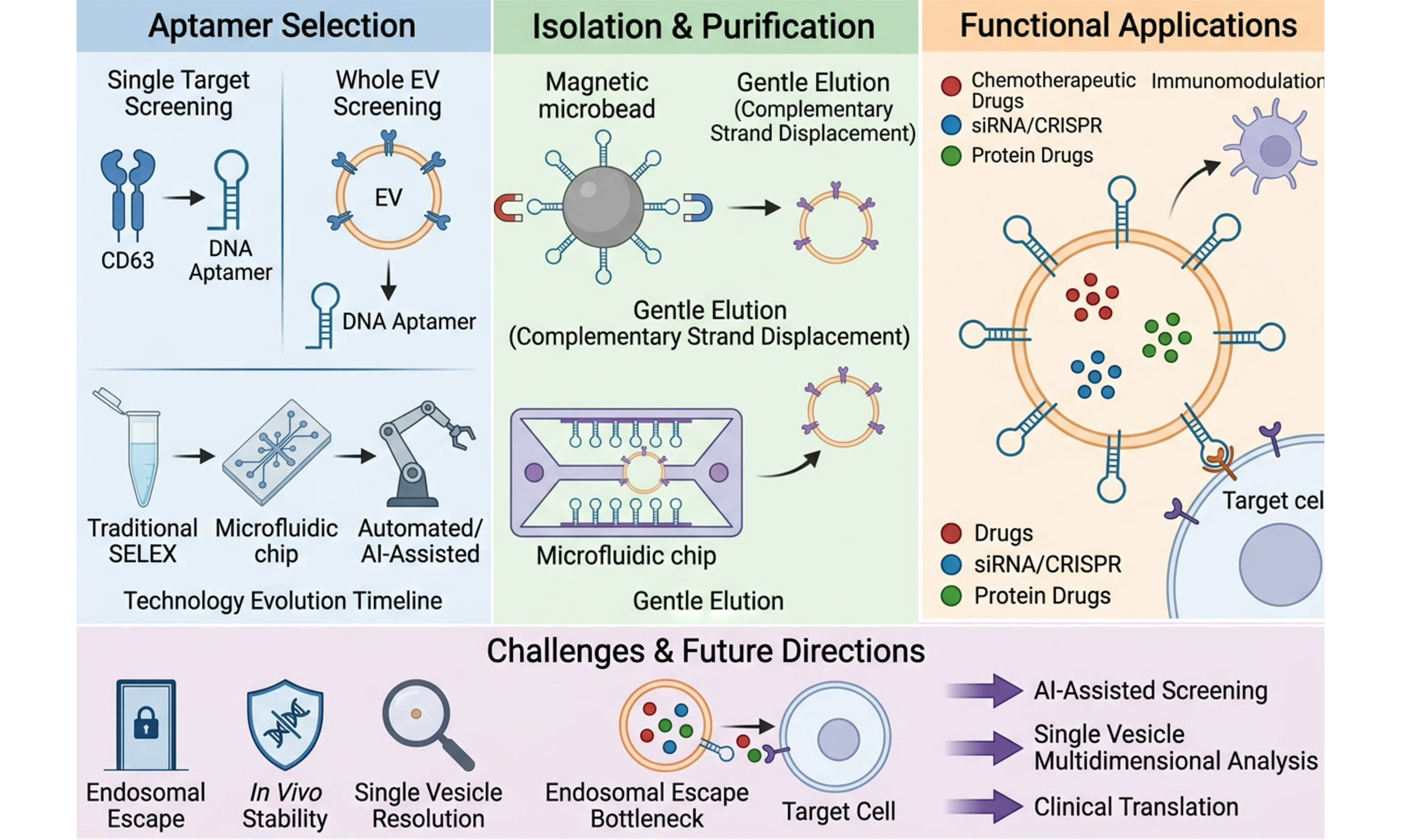
Extracellular vesicles (EVs) are natural mediators of intercellular communication and hold promise for disease diagnosis and drug delivery, yet their intrinsic heterogeneity complicates isolation, molecular profiling, and targeted delivery. Aptamers, owing to their high affinity, specificity, chemical tunability, and scalable synthesis, offer a flexible molecular toolkit to address these challenges. In this review, we highlight recent progress in integrating aptamers into EV research across three interconnected domains: (1) the selection of aptamers against EV-associated surface markers; (2) the development of aptamer-based methods for EV capture and isolation; (3) the use of aptamer-functionalized EVs for targeted drug delivery and immunomodulation. We also discuss outstanding challenges, particularly the need for selection strategies that better preserve native EV epitope conformations and the rational design of multifunctional aptamer architectures. Looking forward, the convergence of aptamer technology with EV biology is expected to enable aptamer-drug conjugates and minimally invasive, dynamic liquid biopsy approaches for major diseases such as cancer, ultimately advancing early diagnosis and precision medicine.
Keywords


 Download PDF
INTRODUCTION
Extracellular vesicles (EVs) are nanoscale lipid bilayer vesicles released by virtually all cell types into biofluids including blood, urine, and saliva[1] (Figure 1, left and right panels: key structural components and biomarkers of exosomes, and their biogenesis via the endosomal system and release pathways). Originally dismissed as cellular debris, EVs are now recognized as key mediators of intercellular communication, shuttling proteins, nucleic acids, and lipids to recipient cells and thereby influencing diverse physiological and pathological processes[2-4]. However, the inherent heterogeneity of EVs, in terms of size, surface marker expression[5], and molecular cargo, poses substantial challenges for their isolation, accurate molecular profiling, and targeted delivery. Conventional isolation methods, including ultracentrifugation, density gradient centrifugation, and size-exclusion chromatography, suffer from low purity, poor reproducibility, and compromised vesicle integrity[6,7]. These limitations underscore the urgent need for affinity-based strategies that enable specific and efficient EV capture.
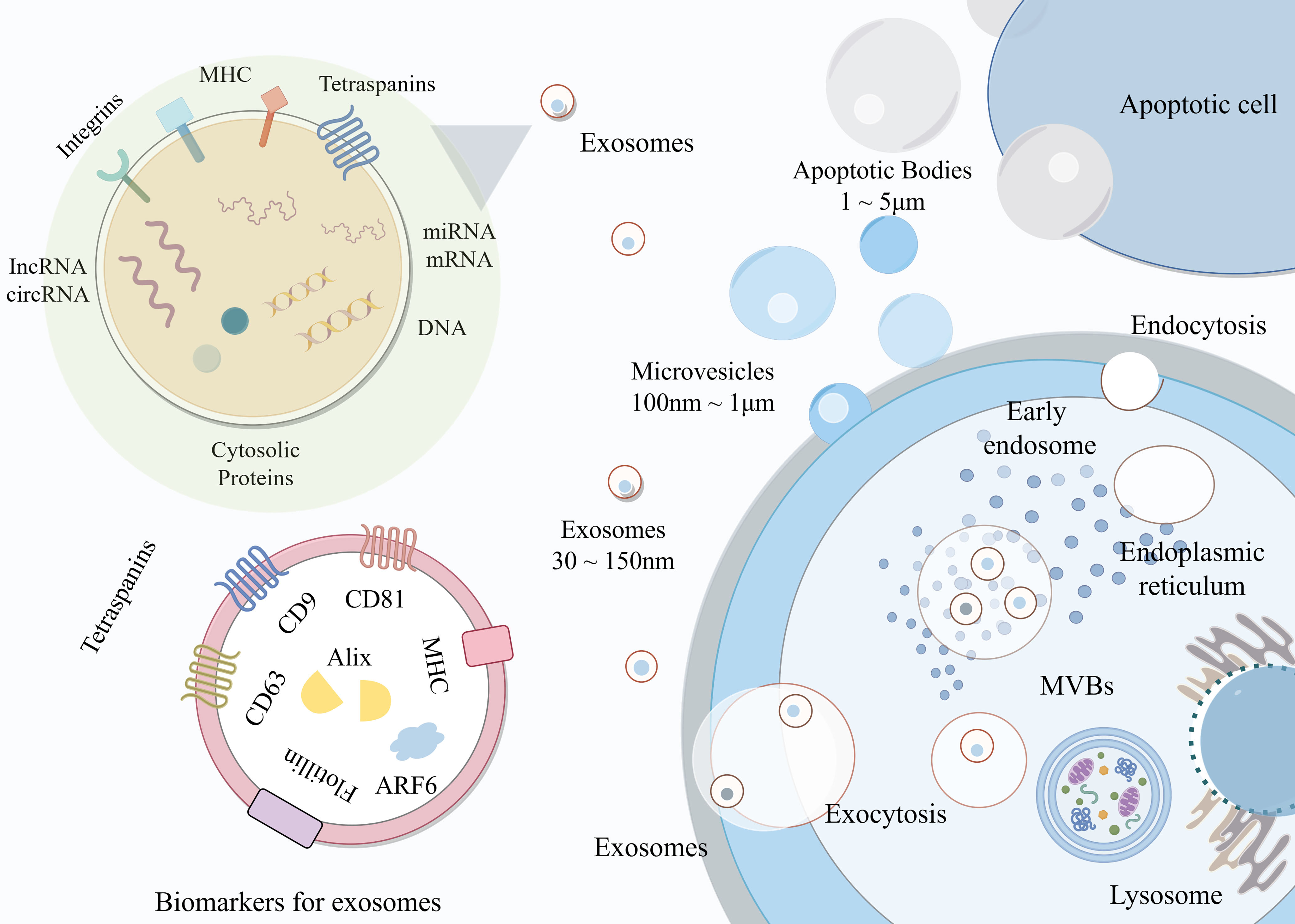
Figure 1. Schematic illustration of EV biogenesis, composition, and secretion. This schematic illustrates the heterogeneity and biogenesis of EVs, which are classified by size into apoptotic bodies, microvesicles, and exosomes. The left panels detail the structural composition of exosomes, highlighting various cargoes. The right panel maps the cellular biogenesis pathway, where endocytosis leads to the formation of early endosomes and subsequent MVBs. MVBs either fuse with the plasma membrane to release exosomes via exocytosis or are directed toward lysosomes for degradation. This figure was drawn with Figdraw and is an original work of the authors. Figdraw ID: WSWOUbbd28. EV: Extracellular vesicle; circRNA: circular RNA; lncRNA: long non-coding RNA; MHC: major histocompatibility complex; miRNA: microRNA; mRNA: messenger RNA; MVB: multivesicular body.
Aptamers, short, single-stranded DNA or RNA molecules generated through systematic evolution of ligands by exponential enrichment (SELEX), have emerged as compelling alternatives to antibodies in EV research[3,8]. Unlike antibodies, aptamers are chemically synthesized, offering batch-to-batch consistency, low immunogenicity, and facile chemical modification with fluorophores, drugs, or anchoring moieties[9-13]. More importantly, aptamers offer three distinct advantages that are particularly relevant to EV studies. First, EV SELEX enables the discovery of novel, disease-specific surface fingerprints without prior knowledge of EV surface epitopes, thereby overcoming the limitation of antibody-based approaches that target only predefined immunogenic proteins[14,15]. Second, aptamer-based EV capture can be reversed under mild conditions using complementary strand displacement, preserving EV integrity and cargo for downstream multi-omics analyses, a unique advantage over the harsh elution conditions (e.g., low pH, high salt, or organic solvents) required for antibody-based capture[16,17]. Third, the scalable, animal-free synthesis of aptamers eliminates the batch variability that has long plagued antibody-based EV diagnostics[11]. A comprehensive comparison of aptamers and antibodies is provided in Supplementary Table 1.
In this review, we examine recent advances in integrating aptamers into EV research across three interconnected domains: the selection of aptamers targeting EV surface markers; the engineering of aptamer-functionalized EVs for targeted drug delivery and immunomodulation; and the development of aptamer-based platforms for EV isolation and purification. We critically assess current challenges, in particular the need for selection strategies that preserve native EV epitope conformations and for the rational design of multifunctional aptamer architectures[18]. Finally, we offer perspectives on how aptamer-EV technologies could enable minimally invasive liquid biopsy strategies and accelerate the clinical translation of EV-based diagnostics and therapeutics.
APTAMER SELECTION STRATEGIES: FROM SINGLE TARGETS TO INTACT EVs
Principles of aptamer selection: single-marker versus whole-EV recognition
Aptamer-based recognition of EVs relies on specific three-dimensional interactions between aptamers and surface molecular features, including proteins, glycans, and lipid moieties[19,20]. Depending on the target form, two complementary strategies have emerged.
The first strategy targets predefined EV surface markers, such as tetraspanins (CD9, CD63, CD81) or disease-associated antigens[17-21]. Aptamers generated against these known epitopes offer well-defined binding sites and facilitate standardized assay development. For instance, slow off-rate modified aptamers (SOMAmers)®-based affinity proteomics has enabled simultaneous profiling of over 1,000 proteins in prostate cancer-derived EVs[22], while artificial intelligence (AI)-assisted SELEX workflows reduced the number of laboratory enrichment cycles from conventional 12-15 to 5-7, as reported in a pooled analysis of 34 AI-integrated SELEX studies[14]. However, single-marker approaches are inherently limited by EV heterogeneity: even EVs from the same parental cell exhibit substantial variations in marker expression, and a single biomarker rarely captures full disease complexity[23,24].
The second, more recent strategy employs intact EVs as selection targets[25,26]. Through positive selection against target EVs and counter-selection against controls, this approach yields aptamers that recognize combinatorial surface fingerprints, encompassing proteins, glycans, and lipids, without requiring prior knowledge of individual epitopes[27]. Such holistic recognition more faithfully reflects the physiological or pathological state of the EV-producing cells. Zhao and colleagues demonstrated this concept by developing a capillary electrophoresis-based, three-step SELEX against intact natural killer cell-derived EVs, obtaining aptamers with a Kd of 27.6 nM within five rounds[28]. Collectively, aptamers thus provide dual functionality, serving as antibody-like probes for known markers or as discovery tools to uncover unknown EV surface features. A schematic representation of the conventional SELEX workflow is shown in Figure 2.
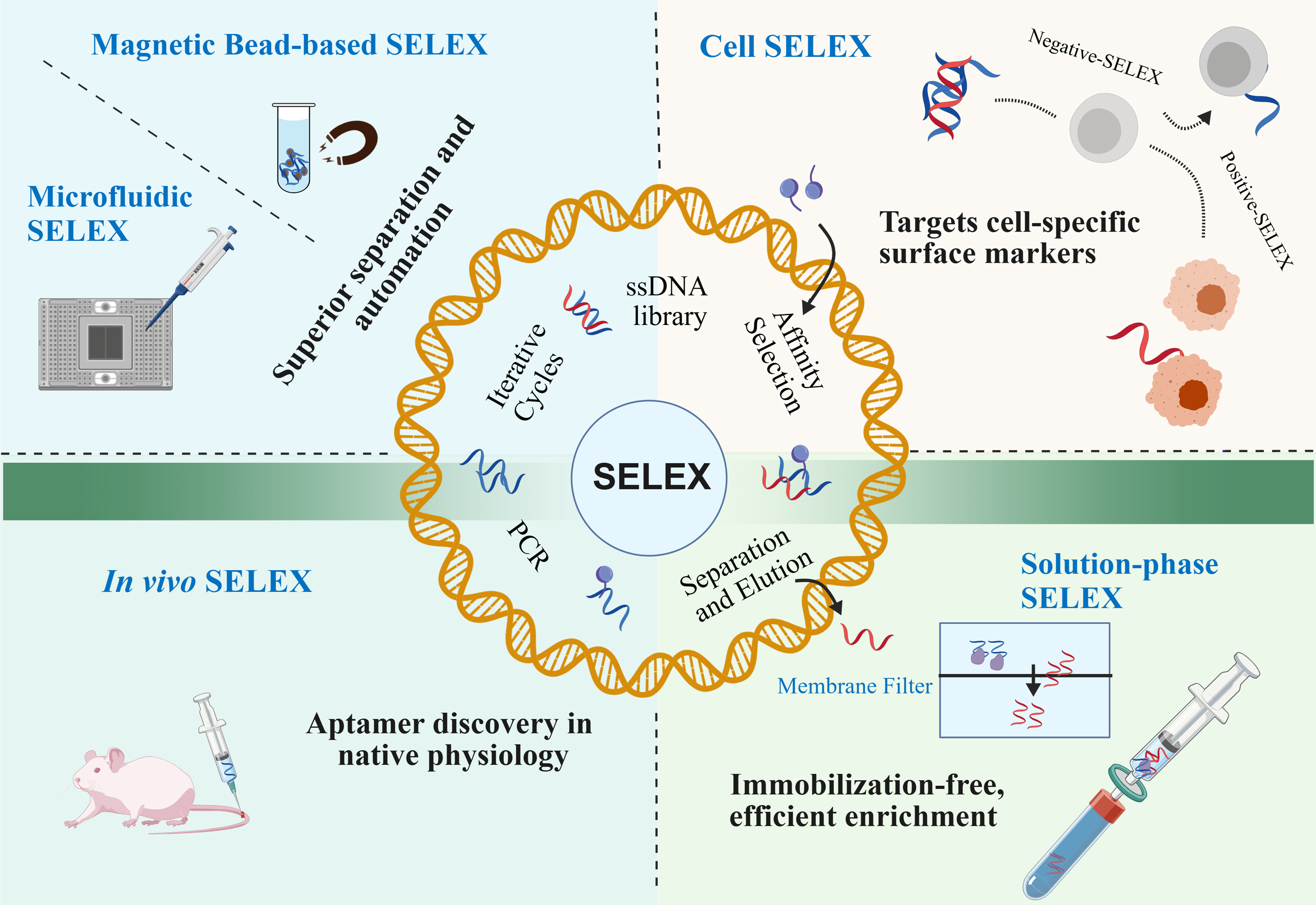
Figure 2. Schematic workflow of SELEX. The central schematic delineates the core iterative SELEX cycle (comprising ssDNA library construction, affinity selection, separation/elution, and PCR amplification), alongside several emerging variants such as Cell SELEX, in vivo SELEX, magnetic bead-based SELEX, and microfluidic SELEX. This figure was drawn with BioGDP and is an original work of the authors. BioGDP Agreement Number:GDP20268V0A4A. PCR: Polymerase chain reaction; SELEX: systematic evolution of ligands by exponential enrichment; ssDNA: single-stranded DNA.
Evolutionary logic of selection technologies
Aptamer selection technologies have evolved through three overlapping phases. The first phase (1990-2010) established conventional SELEX against purified proteins, yet these methods proved ill-suited for EV targets owing to immobilization-induced conformational changes and high sample consumption. The second phase (2010-2020) introduced microfluidics and capillary electrophoresis, substantially reducing selection rounds while preserving native epitope conformations. The third phase (2020-present) is defined by automation, artificial intelligence, and functional selection - integrating robotic liquid handling, real-time monitoring, and machine learning to enable the fully automated selection of intact EVs without operator intervention, with functional activity as a primary selection criterion. Rather than a chronological catalog, we organize the discussion that follows around three core objectives that have driven this evolution: screening efficiency, target complexity, and aptamer functionality.
Improving screening efficiency
The past two decades have seen a paradigm shift from manual, time-intensive SELEX to automated, high-throughput platforms. Traditional SELEX suffers from a high number of selection rounds and long processing times[8,9], as shown in Figure 3A. Integration of SELEX with microfluidics enables efficient aptamer separation, significantly shortening the selection time while minimizing the consumption of biological materials[29,30], as shown in Figure 3B. Capillary electrophoresis SELEX eliminated target immobilization, a critical advantage for preserving native EV epitope conformations, and yielded high-affinity aptamers (Kd = 27.6 nM) in as few as five rounds[28].
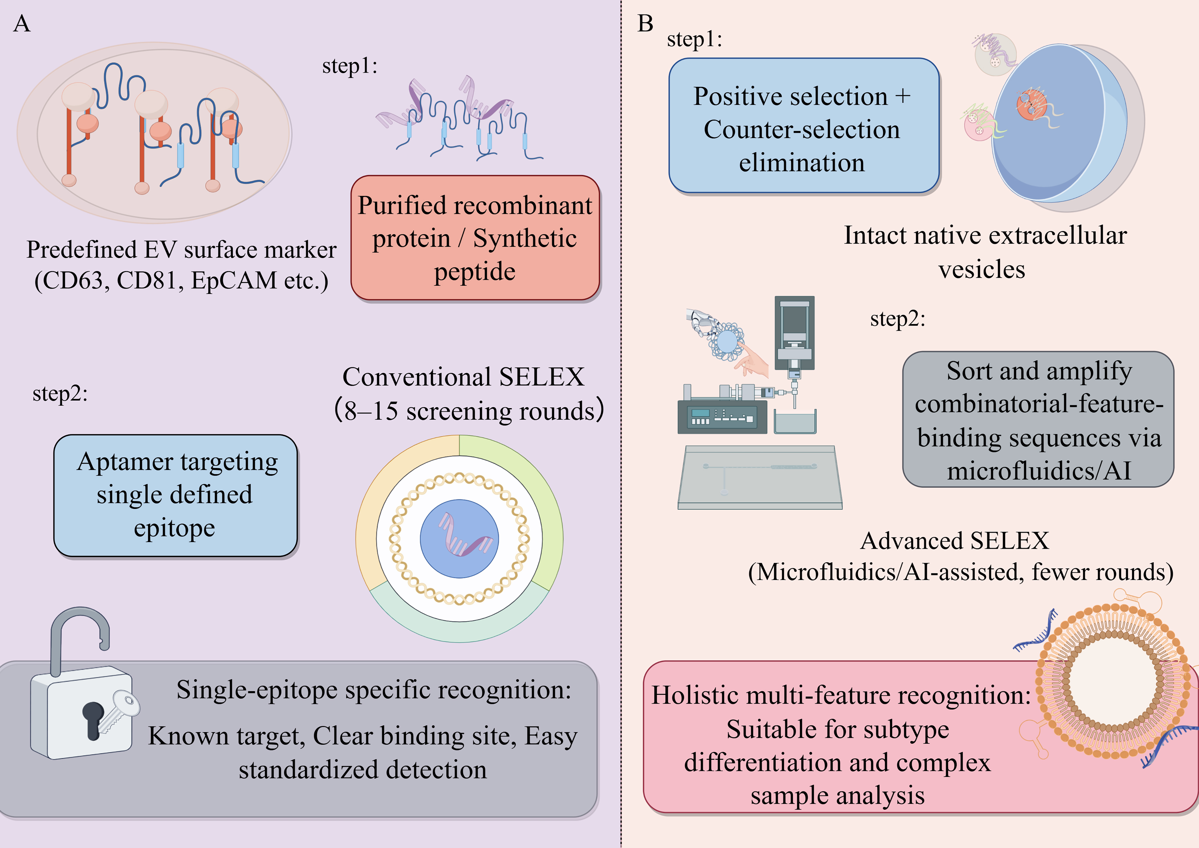
Figure 3. Comparative schematic workflow of SELEX methodologies for EVs. (A) Conventional SELEX strategy. This approach uses purified recombinant proteins or synthetic peptides as targets based on predefined EV surface markers (such as CD63, CD81, EpCAM). It requires 8-15 screening rounds and yields aptamers targeting a single defined epitope. This results in single-epitope-specific recognition, characterized by known targets, clear binding sites, and easy standardized detection; (B) Advanced SELEX strategy. This approach utilizes intact native EVs, incorporating positive selection and counter-selection elimination. It employs microfluidics/AI-assisted systems to sort and amplify combinatorial-feature-binding sequences, requiring fewer rounds. This method achieves holistic multi-feature recognition, making it suitable for subtype differentiation and complex sample analysis. This figure was drawn with Figdraw and is an original work of the authors. Figdraw ID: WYTPRcaf81. AI: Artificial intelligence; EpCAM: epithelial cell adhesion molecule; EV: extracellular vesicle; SELEX: systematic evolution of ligands by exponential enrichment.
Most recently, fully automated platforms integrating microfluidics, robotic liquid handling, real-time quantitative polymerase chain reaction (qPCR) monitoring, and machine learning have achieved the automated, hands-free selection of intact EVs within days[30]. These advances have transformed aptamer discovery from an artisanal craft into an engineering discipline. However, these platforms, alongside existing hybrid approaches that combine robotic handling with microfluidic modalities such as droplet microfluidics or acoustofluidics, still fall short of addressing three essential prerequisites for automated multi-analyte liquid biopsy applications in a single instrument: (1) supporting the processing of large sample volumes; (2) enabling the processing of more than one sample at a time; and (3) providing orthogonal extraction mechanisms as required[31]. Figure 3 presents a comparison of conventional versus advanced SELEX strategies for aptamer selection targeting EVs.
Expanding target complexity
The shift from soluble proteins to intact EVs represents a fundamental expansion in target complexity. Early efforts focused on individual EV surface markers such as epithelial cell adhesion molecule (EpCAM), mucin 1 (MUC1), and CD63[21]; however, such predefined targets cannot resolve EV subset heterogeneity. The breakthrough came with whole-EV SELEX, which treats EVs as complex molecular assemblies[26]. Cell-SELEX against cancer cells followed by counter-selection against normal cells enabled the discovery of aptamers recognizing unknown, disease-specific surface features[32,33]. EDGE-SELEX applied to intact exosomes, together with immobilization-free SELEX that avoids conformational artifacts[26], has further pushed the boundary. Most recently, AI-driven SELEX design has optimized library complexity for multi-epitope recognition on EV surfaces[33], while fully automated platforms now enable programmable, high-throughput selection against intact EVs[29]. Collectively, these advances have shifted the field from targeting purified molecules to confronting physiologically relevant, complex targets.
Enhancing aptamer functionality
Initially regarded as antibody substitutes for detection, aptamers have undergone a functional redefinition: selection criteria now extend beyond binding affinity to encompass biological activity[34,35]. Emerging strategies prioritize aptamers that not only bind with high affinity but block protein-protein interactions, mediate drug internalization, or activate immune signaling[36,37]. Examples include aptamers that block heat shock protein 70 (HSP70)-mediated immunosuppression[38], as well as aptamers that recognize membrane protein targets on exosomes derived from highly metastatic colorectal cancer and directly suppress exosome-induced pro-metastatic activities, such as cell migration, invasion, and angiogenesis[39]. This functional turn shifts the paradigm from binding to intervention, positioning aptamers as therapeutic effectors rather than passive targeting ligands.
Unresolved bottlenecks in aptamer selection for EV research
Despite these advances, several challenges persist
In some reported cases, functional selection may involve a trade-off with binding affinity[34]. For instance, a study using the DNA aptamer GS24 targeting the transferrin receptor revealed that this aptamer exists in two conformations; however, only one of them (A-2F) possesses biological activity. Engineering modifications, such as truncation or mutation, were employed to stabilize the active conformation and enhance its biological activity; nevertheless, the affinity of the truncated variant GS24min was approximately five times lower than that of the parental GS24[40]. However, this trade-off is not a universal rule; whether it occurs depends critically on whether the aptamer’s epitope overlaps with a functional site (e.g., a receptor-binding domain) or a non-functional structural region[34]. Several examples demonstrate that aptamers can maintain both high affinity and full biological activity when the epitope is appropriately positioned[41].
The observed trade-off in EV applications is primarily based on a limited number of systems, such as HSP70[38], and should therefore be considered a cautionary observation rather than an established limitation. Nevertheless, for EV applications where aptamers must simultaneously recognize surface epitopes with high specificity and exert downstream functions (e.g., inhibiting EV-mediated signaling or directing cargo internalization), balancing these two properties remains an important design consideration.
Second, the inherent heterogeneity of EVs, both across and within populations, severely compromises inter-laboratory reproducibility. EVs derived from the same cell type can vary significantly in size, surface marker density, and cargo composition depending on culture conditions, isolation methods, and storage protocols[5]. This batch-to-batch variability means that aptamers selected against one EV preparation may perform poorly against another, even from the same nominal source[23,24]. To address this, the field urgently needs standardized biological and physical reference materials to benchmark aptamer binding affinity and specificity across different laboratories and platforms. Promising candidates include recombinant EVs (rEVs) engineered to mimic native biochemical and biophysical properties for quantitative data normalization, nanoerythrosomes, and engineered vesicles or synthetic liposomes with predefined sizes, refractive indices, and marker/epitope abundances.
Third, concurrent target discovery and functional aptamer selection without prior knowledge of EV surface features remains a technical blind spot. Most current selection strategies either begin with a known target [e.g., CD63, programmed death-ligand 1 (PD-L1)][21] or use whole-EV selection to generate aptamers without immediately knowing their binding targets[32].
However, when an aptamer selected against intact EVs elicits a desired functional effect, such as blocking EV uptake or neutralizing a pathological activity, identifying its cognate surface epitope becomes a major bottleneck. Reverse engineering the target from a functional aptamer is technically demanding and time-consuming, typically requiring immunoprecipitation, mass spectrometry, or clustered regularly interspaced short palindromic repeats (CRISPR)-based validation. Until this blind spot is resolved, the systematic discovery of novel EV surface biomarkers that are both therapeutically actionable and diagnostically useful will remain constrained.
APTAMER-MEDIATED EV ISOLATION AND PURIFICATION
Principles and key advantages of aptamer-based isolation
High-purity EV isolation is a prerequisite for downstream molecular analysis and functional studies. Conventional methods, such as ultracentrifugation, density gradient centrifugation, size-exclusion chromatography, and polymer precipitation, are hampered by low purity, long processing times, heavy equipment dependence, and an inability to resolve EV subtypes[6,7,42]. Aptamer-based affinity capture overcomes these limitations through two distinctive advantages.
First, aptamers offer high specificity for EV surface epitopes, including those with low abundance or post-translational modifications[43,44]. Second, and uniquely, aptamers enable mild, non-destructive elution via complementary strand displacement, preserving EV integrity and cargo for downstream multi-omics analyses[45,46]. This contrasts sharply with antibody-based capture, which requires harsh conditions (low pH, high salt, or organic solvents) that compromise vesicle structure.
Evolution of technology platforms for aptamer-based EV isolation
Magnetic bead-based platforms
Magnetic separation is the most established aptamer-based platform for EV isolation. Biotin-labeled aptamers are immobilized on streptavidin-coated magnetic beads[47,48], enabling rapid EV capture under an external magnetic field. At the optimal aptamer concentration of 1 μM, the capture efficiency reaches 80%, significantly higher than the 65% achieved by conventional antibody-functionalized Fe3O4 beads[48]. Single-target magnetic separation has since been applied to various disease-associated EVs[49].
Multitarget magnetic separation further improves specificity and coverage[50,51]: dual-aptamer systems integrated with graphene field-effect transistors permit label-free detection of cancer-derived vesicles[42], while triple-aptamer electrochemical approaches simultaneously recognize multiple EV surface epitopes[52]. Critically, complementary strand displacement and deoxyribonuclease I cleavage have been optimized for non-destructive elution, both of which preserve EV morphology and membrane protein integrity[53,54].
Microfluidic chip platforms
Microfluidics integrates EV isolation into miniaturized chip formats, substantially reducing sample consumption and processing time[55]. Aptamer-based microfluidic devices enable target-specific EV capture with efficiencies of 107-108 particles/mL[56], while select filtration-based platforms enable direct EV isolation from whole blood without pre-processing[57]. Magnetic microfluidics further enhances isolation efficiency by combining magnetic separation with on-chip flow control, completing isolation within 1 h[58,59].
However, substantial challenges remain. Wide variability in chip geometry, flow rate, and surface chemistry across laboratories hinders cross-study comparison, and the lack of standardized reference samples for calibration impedes clinical translation.
Smart responsive materials
Smart responsive materials enable dynamic control over EV capture and release. Thermosensitive aptamer-conjugated materials achieve > 60% recovery rate through temperature switching[21]. Photoactivatable magnetic beads offer spatiotemporal precision[60]. DNA hydrogels incorporating CRISPR/CRISPR-associated protein 12a (Cas12a) respond to EV recognition through conformational changes, enabling signal-triggered release. Moreover, these hydrogels possess a high encapsulation capacity, with a maximum of 7.6 × 10⁷ exosomes per 50 μL of hydrogel, which facilitates efficient EV loading for downstream analysis[61,62]; in this system, EV binding induces a conformational change in the hydrogel, activating Cas12a’s trans-cleavage activity, which then releases a reporter or triggers cargo liberation. Molecularly imprinted polymers combined with aptamers can recognize glycosylation patterns, a capability beyond conventional aptamers[63].
Although these platforms have expanded the toolbox from passive adsorption to active regulation, most remain confined to benchtop operation[64]; the lack of integrated automated liquid handling and external temperature/light control modules continues to impede their routine use.
Integrated multidimensional platforms
Integrated platforms that combine isolation with downstream analysis enable sample-to-answer workflows. The CLCtips platform, for instance, integrates aptamer-mediated capture and on-chip enzymatic digestion for trace EV proteomics, identifying over 500 proteins from minimal sample volumes[65]. Microfluidic chips coupled with mass spectrometry or next-generation sequencing permit multi-omics profiling from single isolation runs[66-68], while proximity hybridization-mediated FRET (Fluorescence Resonance Energy Transfer) assays achieve high-specificity EV detection without separate isolation steps[69].
Key bottlenecks for integrated platforms include multi-step compatibility (capture, lysis, digestion, analysis), the purity-sensitivity trade-off, sample throughput, and automation costs.
Current bottlenecks for aptamer-based EV isolation platforms
Magnetic bead-based platforms offer a leading combination of maturity and ease of use, with consistently outstanding capture efficiency[47-49]. Microfluidic platforms deliver the best combination of speed and purity[58], but microfluidic and acoustofluidic methods also exhibit limited selectivity and frequently yield impure isolated samples. Immunoaffinity-based approaches can enhance specificity through molecular recognition; however, their applicability to multiplex separation remains constrained[70]. Smart responsive materials excel in controllable release and reusability[21], yet their dependence on external temperature or light control modules complicates workflow integration[71,72]. Integrated multidimensional platforms minimize sample handling by combining isolation with downstream analysis[66], but the purity-sensitivity trade-off remains unresolved[73,74]. Collectively, no single platform meets all clinical requirements. Every method involves intrinsic compromises between recovery efficiency, purity, scalability, and the maintenance of vesicular integrity[75-77]. Moreover, inter-laboratory variability remains a significant impediment, as even slight discrepancies in isolation or handling procedures can markedly affect exosomal yield, molecular composition, and downstream biomarker measurements[77]. In addition to these trade-offs, isolation time has emerged as a critically important parameter, particularly for clinical applications where rapid turnaround is essential for timely diagnosis and for preserving the integrity of labile RNA and protein cargoes. Extended protocols not only reduce sample throughput but also increase the risk of degradation and batch-to-batch variation, hampering translation into routine practice. To clarify this key metric, Table 1 summarizes the isolation times of major aptamer-based EV isolation platforms.
Time comparison of aptamer-based EV isolation platforms
| Technology platform | Carrier | Time distribution | Ref. |
| Magnetic beads | mesoporous Fe3O4@Au nanoparticles conjugated with CD63 DNA aptamer | Capture incubation: 60 min; Elution: 20 min | [48] |
| Magnetic microfluidic | Tentacle-like magnetic microspheres conjugated with CD63 aptamer | EV enrichment: 20 min; Exonuclease-I-mediated release: 30 min | [58] |
| DNA-based hydrogel | RCA-synthesized ultralong DNA chains containing polyvalent CD63 aptamer | Capture incubation: 30 min; Hydrogel formation incubation 30 min | [61] |
| Smart responsive materials | Thermosensitive copolymer PNB grafted with CD63 aptamer | Capture incubation: 120 min; Complementary-sequence elution: 15 min | [21] |
APPLICATION OF APTAMER-FUNCTIONALIZED EVS IN DRUG DELIVERY AND IMMUNOMODULATION
Aptamers offer high affinity, low immunogenicity, facile chemical modification, and scalable synthesis, making them ideal targeting moieties for EV functionalization[78,79]. This section examines two major application domains: targeted drug delivery and immunomodulation. Within drug delivery, we organize by cargo type, namely chemotherapeutics, nucleic acids, and proteins, while highlighting a critical bottleneck common to all: endosomal escape.
Aptamer-mediated targeted drug delivery using EVs
The core strategy involves anchoring aptamers onto EV surfaces to enable specific recognition of diseased cells[80,81]. Three functional modules, including targeting (aptamer), carrier (EV), and cargo (therapeutic), can be combined in various configurations.
Targeted delivery of chemotherapeutic agents
Aptamer-functionalized EVs are engineered to serve as a safe and effective targeted drug delivery system for specific anatomical sites, improving long-term persistence and boosting the safety and efficacy of chemotherapy treatments[82]. Two internalization routes have been exploited: receptor-mediated endocytosis and membrane fusion. Most strategies rely on clathrin- or caveolae-mediated endocytosis initiated by aptamer-receptor binding[83,84]. For example, the nucleolin-targeting AS1411 aptamer delivers doxorubicin via clathrin-mediated endocytosis[82], and mesenchymal stem cell-derived EVs combine intrinsic homing with aptamer recognition for dual targeting[85]. A disulfide linker between the aptamer and EV enables glutathione-triggered drug release in the cytoplasm[86,87], while raloxifene-conjugated systems use lysosomal targeting to induce both autophagy and apoptosis, a dual mechanism with the potential to circumvent chemoresistance[88]. Representative examples of receptor-mediated endocytosis pathways are summarized in Supplementary Table 2. A systematic schematic overview of all four major internalization routes (clathrin-mediated endocytosis, caveolin-mediated endocytosis, macropinocytosis, and direct membrane fusion) and their representative applications in aptamer-EV delivery systems is provided in Figure 4.
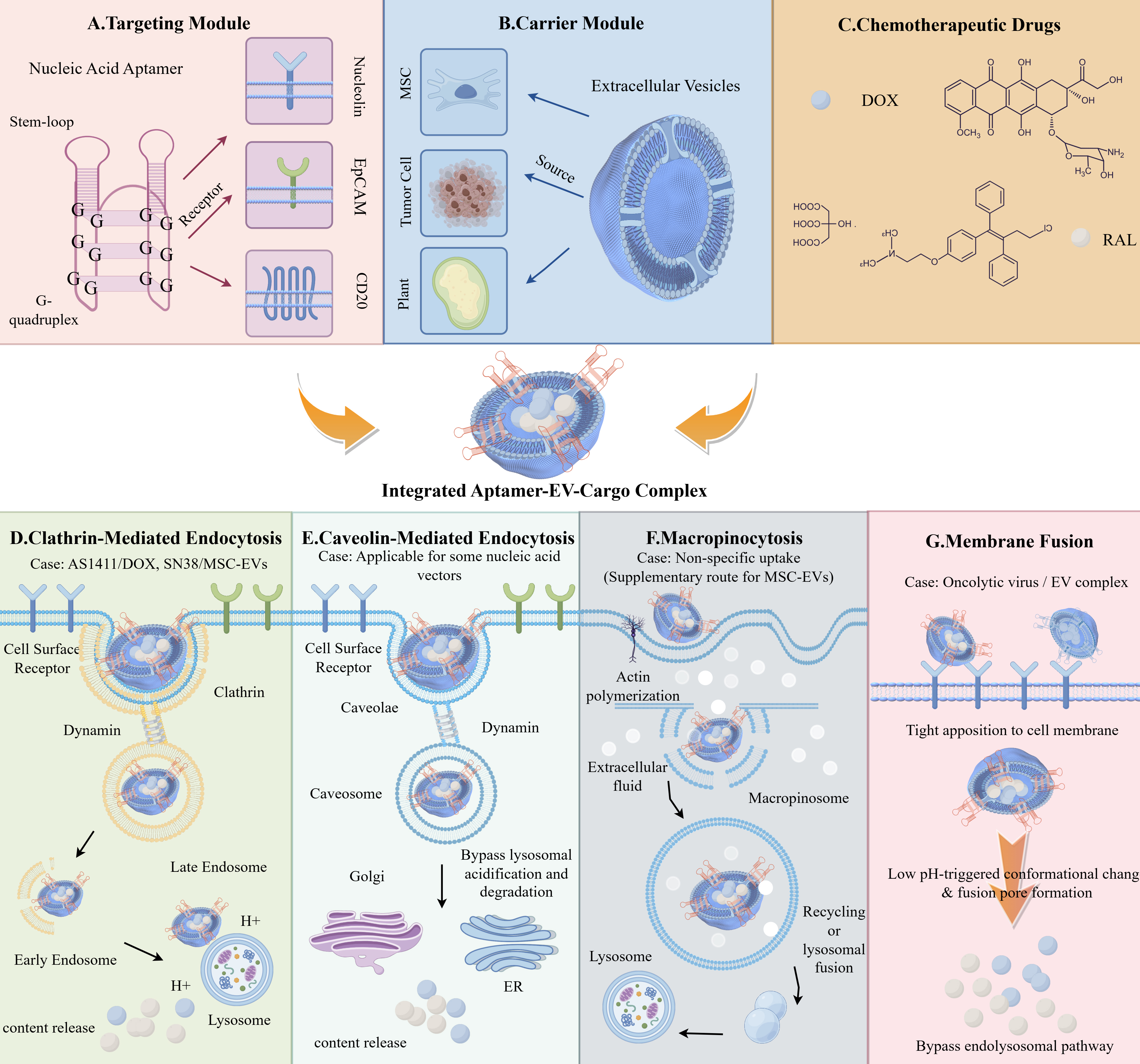
Figure 4. Modular design of aptamer-functionalized EV-based chemotherapeutic delivery systems. Upper schematic: This schematic illustrates the rational design of an integrated aptamer-EV-drug delivery system, which is assembled from three functional modules: (A) the targeting module, comprising nucleic acid aptamers with characteristic secondary structures (stem-loop, G-quadruplex) that bind to specific cell surface receptors (e.g., nucleolin, EpCAM, CD20); (B) the carrier module, consisting of EVs derived from multiple sources including MSCs, tumor cells, and plants; and (C) the chemotherapeutic drug module, represented by DOX and RAL. These three modules are conjugated to form the integrated Aptamer-EV-Cargo complex for targeted delivery. Lower subpanels (D-G): The lower portion delineates four distinct cellular internalization routes for the integrated complex, demonstrating versatile pathways for intracellular cargo release and potential avoidance of lysosomal degradation. (D) Clathrin-mediated endocytosis pathway, which is the primary route for AS1411/DOX and SN38/MSC-EVs internalization. (E) Caveolae-mediated endocytosis, suitable for nucleic acid vectors, which bypasses lysosomal acidification and degradation. (F) Macropinocytosis pathway, representing a non-specific uptake route that serves as a supplementary pathway for MSC-EVs. (G) Membrane fusion pathway, utilized by oncolytic viruses/EV complexes, involving tight apposition and low pH-triggered conformational changes to bypass the endolysosomal pathway. This figure was drawn with Figdraw and is an original work of the authors. Figdraw ID: TRSPAdd884. DOX: Doxorubicin; EpCAM: epithelial cell adhesion molecule; EV: extracellular vesicle; MSC: mesenchymal stem cell; RAL: raloxifene; SN38: irinotecan active metabolite SN38.
Despite these advances, all receptor-mediated pathways are constrained by inefficient endosomal escape[89-91]: only a minor fraction of internalized cargo reaches the cytosol[92]. This limitation is tolerable for potent chemotherapeutics but becomes critical for biologics. To demonstrate aptamer-mediated targeted docking, researchers developed a multifunctional exosome system, ExomiR-CVB3/DoxApt, comprising oncolytic coxsackievirus B3-derived exosomes[93], a surface-conjugated AS1411 aptamer for nucleolin recognition, and encapsulated doxorubicin. In the acidic tumor microenvironment, aptamer-mediated nucleolin anchoring coincides with a sharp pH-dependent increase in drug release, resulting in localized drug deposition at the target cell surface[94]. This design provides mechanistic evidence for aptamer-assisted targeting: gel shift assays confirmed stable aptamer conjugation, and the differential release profile indicates preferential cargo unloading following aptamer-guided localization to the acidic tumor milieu. Nevertheless, these data offer indirect support for targeted docking; direct cell-binding or uptake studies are still required to fully validate the targeting advantage, and the potential off-target immunogenicity of the viral backbone must be carefully managed.
Targeted delivery of nucleic acid drugs
Target selection for aptamer-mediated EV delivery generally adheres to two criteria: (i) overexpressed surface biomarkers unique to pathological tissues [e.g., EpCAM, human epidermal growth factor receptor 2 (HER2), nucleolin, CD20]; and (ii) receptors capable of mediating robust internalization (e.g., transferrin receptor). Whole-cell SELEX offers an alternative route, enabling isolation of functional aptamers without prior target identification, provided they support intracellular uptake.
Nucleic acid therapeutics - small interfering RNA (siRNA), messenger RNA (mRNA), circular RNA (circRNA), and CRISPR-Cas9 - all require cytosolic delivery for activity. Aptamers serve a dual role, directing cell-specific uptake and, in certain designs, facilitating endosomal escape. For RNA interference, aptamer-displayed EVs have been validated for circRNA delivery to cancer cells[95], and CL4 aptamer-modified EVs delivering long non-coding RNA (lncRNA) DARS-AS1 siRNA have been shown to reverse chemoresistance in triple-negative breast cancer[96]. For mRNA delivery, hepatocyte-targeted systems have been developed for familial hypercholesterolemia[97]; circular RNA and microRNA (miRNA) sponge delivery via aptamer-EV platforms has also been demonstrated[98,99].
The most advanced application is CRISPR-Cas9 delivery. Elsharkasy et al. devised a modular strategy in which aptamers mediate ribonucleoprotein (RNP) loading into EVs, with ultraviolet (UV)-triggered release upon endocytosis[100]; Hegeman et al. further refined aptamer affinity to enhance delivery precision[101]; and Xu et al. engineered a reactive oxygen species (ROS)-responsive, aptamer-conjugated EV system for combined oncolytic virus OH2 and anti-PD-L1 therapy in prostate cancer[102].
The endosomal escape bottleneck is most severe for these cargoes. Whereas a few siRNA copies per cell can achieve potent gene silencing, mRNA and CRISPR-Cas9 demand substantially higher cytosolic concentrations[99,100]. Current efforts to optimize escape through aptamer design - surface charge modulation or minor conformational adjustments - remain largely empirical and insufficient. Transformative progress will likely depend on equipping EVs with viral fusion machinery.
Targeted delivery of protein therapeutics
Protein therapeutics (molecular weight > 10 kDa, fragile tertiary structures) impose additional demands: protection during delivery[103,104], controlled release, and evasion of ubiquitin-proteasome degradation. Aptamer-EV systems have addressed these challenges through three functional paradigms.
Functional antagonism: the aptamer itself acts as the effector. Gobbo et al. designed a peptide aptamer targeting HSP70 on tumor EVs, thereby blocking Toll-like receptor 2 (TLR2)/TLR4-mediated dendritic cell suppression[38]; Quintavalle et al. reported an aptamer against gremlin-1 that disrupts tumor-stroma crosstalk in breast cancer[105].
Growth factor delivery: Luo et al. conjugated aptamers to bone marrow stromal cell EVs - directing them to hydroxyapatite or bone sialoprotein - while encapsulating bone morphogenetic protein 2 (BMP-2), thus shielding the growth factor from circulating proteases and preserving its bioactivity[106].
Gene editor delivery: as described above, aptamer-EV systems for CRISPR-Cas9 RNP delivery represent the leading edge of this field[101].
Aptamer-mediated immunomodulation using EVs
Immunomodulation using aptamer-functionalized EVs spans three areas: cancer immunotherapy, immune cell engineering, and therapy monitoring.
Cancer immunotherapy
Three strategies targeting different nodes of the tumor-immune axis have emerged. The first strategy focuses on blocking immunosuppressive signals. Aptamers against HSP70 or PD-L1 on tumor EVs prevent these molecules from engaging immune receptors[38,107], thereby restoring dendritic cell maturation and T cell activation. The second strategy involves delivering immunostimulatory agents. EpCAM aptamer-modified bacterial EVs serve as tumor vaccines, combining targeted delivery with natural adjuvant activity to enhance antitumor immunity[108]. The third strategy aims at eliminating suppressive EVs themselves. Aptamer-guided capture removes pro-inflammatory or immunosuppressive EV subsets, as demonstrated in allergic airway inflammation, and this approach holds promise for reshaping the tumor microenvironment[109]. Together, these three strategies illustrate the versatility of aptamer-EV platforms in reprogramming immune responses against cancer.
Immune cell engineering
Immune cell-derived EVs naturally carry major histocompatibility complex (MHC) molecules and co-stimulatory signals, making them attractive platforms for immunomodulation. However, their therapeutic utility is limited by poor targeting efficiency and heterogeneous production. Aptamer modification addresses the first limitation by enhancing the tumor-homing capacity of immune cell-derived EVs, enabling their accumulation at disease sites with higher precision[109,110]. To tackle the second, microfluidic strategies enable size-based separation of T cell receptor (TCR)-CD3 EV subsets, ensuring manufacturing consistency and reproducible therapeutic outcomes[23]. Beyond oncology[111], this engineering approach has shown promise in inflammatory diseases: aptamer-functionalized EVs loaded on biomimetic periosteum can recognize nerve growth factor (NGF) released from injured neurons, delivering anti-inflammatory cytokines and neurotrophic factors to promote tissue repair[112]. Collectively, these strategies demonstrate that aptamer-mediated engineering can simultaneously improve targeting accuracy and manufacturing consistency of immune cell-derived EVs, addressing two key barriers to their clinical translation.
Immunotherapy monitoring
Real-time monitoring of immune checkpoint expression on circulating EVs is essential for evaluating patient responses to immunotherapy and guiding treatment decisions. Aptamers meet this need by enabling specific capture and high-sensitivity quantification of immune checkpoint molecules on circulating EVs[113]. For example, He et al. developed a wash-free, homogeneous assay based on an aptamer-cholesterol-anchored DNAzyme assembly, simplifying the workflow without sacrificing accuracy[114]. In another example, Cong et al. constructed a microfluidic device-based extracorporeal circulation system that combines CD63 aptamer-modified magnetic nanospheres for small extracellular vesicle (sEV) capture and a PD-L1-specific trigger aptamer for immune checkpoint recognition, coupled with hybridization chain reaction (HCR) for fluorescence signal amplification; this platform enables continuous in vivo monitoring of PD-L1+ sEVs during tumor progression, reflecting tumor burden and distinguishing early-stage from advanced disease, thus providing real-time feedback for immunotherapy management[55]. In addition, a range of signal transduction platforms have been adapted for aptamer-based EV detection, including SERS (Surface-Enhanced Raman Spectroscopy) probes[115], quantum dot nanospheres integrated with machine learning[116], ellipsometry, and thermophoretic assays[117]. All these platforms share a common three-step cascade - aptamer capture, signal amplification, and quantitative readout - with the principal differences residing in the signal transduction module and achievable sensitivity, entailing trade-offs among complexity, cost, and detection limits. A detailed comparison of these immune monitoring platforms is provided in Supplementary Table 3.
Tissue regeneration and other applications
Aptamer-EV systems have been applied to bone, nerve, and vascular regeneration. Luo et al. pioneered bone targeting by functionalizing bone marrow stromal cell (BMSC)-derived EVs with hydroxyapatite-binding aptamers and loading them with BMP-2[106], while Su et al. achieved combined neural, vascular, and bone repair via NGF targeting and c-Jun N-terminal kinase 3 (JNK3) mitogen-activated protein kinase (MAPK) pathway activation[112].
In neurodegenerative diseases, Ren et al. designed DNA aptamers against α-synuclein aggregates, enabling EV-mediated delivery of chaperones or autophagy inducers to attenuate pathology in Parkinson’s disease models[118]; Du et al. constructed multi-target engineered hybrid EVs bearing amyloid beta (Aβ)-specific aptamers for both oligomer neutralization and anti-inflammatory miRNA delivery[119].
For anti-infection applications, Zhang et al. employed marine-derived EVs with bacterial-targeting aptamers for infected bone defect repair[120], and Cui et al. developed a glycosylated extracellular vesicle-like receptor (termed GlycoEVLR) that enables rapid detection of viral antigens[121].
Collectively, tissue regeneration and anti-infection applications constitute emerging frontiers where aptamer-EV systems offer distinct advantages, notably the capacity to target specific cell types or anatomical sites without requiring endosomal escape for therapeutic activity. Unlike intracellular drug delivery, where cytosolic cargo release is essential, tissue regeneration frequently benefits from sustained surface signaling or paracrine effects[122], thereby mitigating the endosomal escape bottleneck. This distinction indicates that aptamer-EV technologies may achieve earlier clinical translation in regenerative medicine than in intracellular drug delivery.
Key limitations of aptamer-engineered EVs for drug delivery and disease treatment
Despite the promising preclinical results discussed above, several critical limitations continue to impede the clinical translation of aptamer-engineered EVs. Three issues warrant emphasis. First, endosomal escape remains the dominant bottleneck for nucleic acid and protein cargoes[89]; even when aptamers mediate efficient cellular internalization, the vast majority of payloads are retained within endolysosomal compartments and fail to reach their cytosolic or nuclear targets. Second, the in vivo stability of aptamer-EV conjugates under physiological conditions - including exposure to serum nucleases, shear stress, and protein adsorption - is rarely systematically evaluated[55], leaving their true pharmacokinetic profiles poorly defined. Third, manufacturing consistency[23], spanning aptamer-EV conjugation efficiency and batch-to-batch EV homogeneity, remains unstandardized, posing significant obstacles to regulatory approval and scalable production. Future progress will depend on engineering active endosomal escape mechanisms (e.g., viral fusogens, pH-sensitive peptides), rigorous pharmacokinetic and pharmacodynamic characterization in relevant animal models, and developing scalable, Good Manufacturing Practice (GMP)-compliant production protocols[116].
REMAINING HURDLES AND EMERGING SOLUTIONS FOR APTAMER-EV TECHNOLOGIES
A schematic representation of the core challenges and future directions for aptamer-EV technologies is shown in Figure 5.
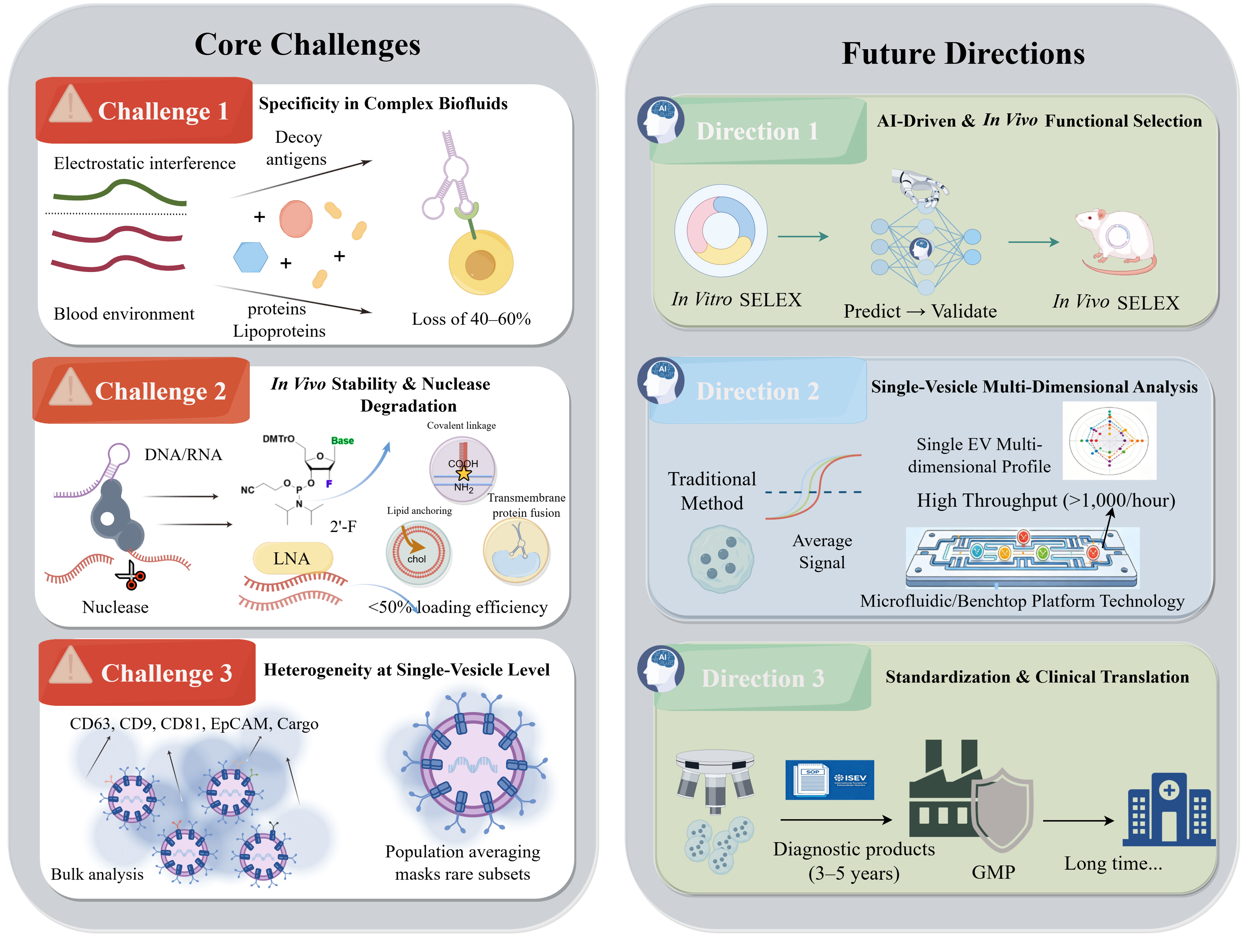
Figure 5. Core challenges and future directions in aptamer-EV technology. This schematic diagram summarizes the critical challenges and prospective future directions in EV-based research and therapeutics. This figure was drawn with Figdraw and is an original work of the authors. Figdraw ID: TRYAU8378a. AI: Artificial intelligence; EpCAM: epithelial cell adhesion molecule; EV: extracellular vesicle; GMP: Good Manufacturing Practice; ISEV: International Society for Extracellular Vesicles; LNA: locked nucleic acid; RNA: ribonucleic acid; SELEX: systematic evolution of ligands by exponential enrichment.
Core challenges
Specificity in complex biological fluids: Biofluids contain abundant proteins, lipoproteins, and free nucleic acids that interfere with aptamer-EV binding. Non-specific adsorption, driven by electrostatic interactions between negatively charged aptamers and cationic proteins, can reduce effective aptamer concentration within 30 min in serum. Soluble antigens may act as decoys, consuming functional aptamers. Chemical modifications (e.g., 2’-O-methyl, locked nucleic acids) and inert carrier surfaces (e.g., PEGylated beads) partially mitigate these issues but may alter aptamer conformation[7,123].
In vivo stability and nuclease degradation: The serum half-lives of unmodified DNA and RNA are 30-60 min and several seconds, respectively[10]. To enhance the serum stability of DNA-and RNA-based aptamers, various chemical modifications have been developed, such as 2’-aminopyrimidine, 2’-O-methylribonucleoside, and 2’-fluoropyrimidine. But excessive modification often reduces binding affinity. EVs themselves offer a solution as natural delivery vesicles: loading aptamers into EV lumens protects them from enzymatic degradation[79]. However, loading efficiency remains suboptimal and carrier-induced immunogenicity is a concern[94].
Heterogeneity resolution at the single-vesicle level: Because EVs from the same parent cell can vary markedly in surface marker expression and cargo, bulk measurements inevitably average out subset heterogeneity. Current aptamer-based platforms operate at the population level[31], masking functional differences between EV subpopulations[30] that may hold distinct diagnostic or therapeutic significance. For instance, only a minor fraction of tumor-derived EVs may carry metastasis-promoting cargo, yet population-level analysis cannot resolve this subset from the predominantly benign background. Single-vesicle analysis, enabled by microfluidics coupled with single-molecule detection or high-resolution imaging[46], remains at an early stage for aptamer-based platforms. Key technical hurdles include achieving sufficient signal amplification to detect low-abundance surface markers on individual vesicles (each EV carries only tens to hundreds of copies of a given protein) and maintaining high throughput[59] while attaining single-vesicle resolution.
Future directions
AI-driven and in vivo functional selection: Deep generative models and machine learning can predict aptamer-target interactions, optimize library design, and reduce selection cycles from 12-15 to 5-7[14]. Integrating multimodal data encompassing sequence, secondary/tertiary structure, chemical modifications, and target functionality will enable first-principles performance prediction, potentially reducing experimental screening to a validation step. Beyond the test tube, in vivo SELEX - direct selection in animal models followed by high-throughput sequencing, promises aptamers that withstand nuclease degradation and delivery barriers in native physiological environments. A key challenge remains: coupling in vivo selection with functional readouts (e.g., therapeutic efficacy rather than mere binding).
Single-vesicle multi-dimensional analysis: The convergence of microfluidics, single-molecule fluorescence, and aptamer-based recognition will enable simultaneous profiling of proteins, nucleic acids, and lipids on individual EVs[66,67]. This capability will permit deconvolution of EV subset heterogeneity and identification of rare, disease-secretory subpopulations, moving beyond population averages. However, single-vesicle analysis remains technically demanding: current methods offer limited throughput and require specialized instrumentation[124]. The near-term goal should be to develop benchtop-compatible platforms that can profile thousands of individual EVs within clinically relevant timeframes.
Standardization and clinical translation: Establishing standardized operating procedures and quality control systems for aptamer-EV technologies is an urgent priority[125,126]. Key parameters, aptamer sequence integrity, modification site occupancy, EV size distribution, and marker expression must be defined and benchmarked against reference materials such as those recently proposed by the International Society for EVs. Large-scale, multi-center prospective studies are needed to validate diagnostic, prognostic, and predictive performance[127]. Plant-derived EVs offer a scalable manufacturing platform[128], but batch consistency and Good Manufacturing Practice compliance require further development.
DECLARATIONS
Acknowledgments
This Graphical Abstract (GA) was created using BioGDP and constitutes an original work of the authors. BioGDP Agreement Number: GDP2026WQAEEJ.
Authors’ contributions
Wrote and edited the manuscript: Li X, Zhang Y
Contributed to visualization, investigation, and conceptualization: Zhang D, Pengsakul T, Qiao Y
Performed review and editing, acquired funding, and participated in conceptualization: Huang L, Zheng J
All authors discussed the results and commented on the manuscript.
Availability of data and materials
Not applicable.
AI and AI-assisted tools statement
During the preparation of this manuscript, the AI tool ChatGPT (version GPT-4, released 2023-03-14) was used solely for language editing. The tool did not influence the study design, data collection, analysis, interpretation, or the scientific content of the work. All authors take full responsibility for the accuracy, integrity, and final content of the manuscript.
Financial support and sponsorship
This work was supported by the Ministry of Science and Technology of the People’s Republic of China (No. 2025YFD2400301), the National Natural Science Foundation of China (No. 32573562), the National Natural Science Foundation of Fujian Province (No. 2026J01314324), the Project of Industry-University Cooperation for Universities in Fujian Province (No. 2024N5006), the Fujian Provincial Spark Project (No. 2024S0008), the Xiamen Municipal Bureau of Science and Technology (No. 3502Z202571006), and the Natural Science Foundation of Xiamen City (No. 3502Z202573062).
Conflicts of interest
All authors declared that there are no conflicts of interest.
Ethical approval and consent to participate
Not applicable.
Consent for publication
Not applicable.
Copyright
© The Author(s) 2026.
Supplementary Materials
REFERENCES
1. Kalluri R, LeBleu VS. The biology, function, and biomedical applications of exosomes. Science. 2020;367:eaau6977.
2. Liu YJ, Wang C. A review of the regulatory mechanisms of extracellular vesicles-mediated intercellular communication. Cell Commun Signal. 2023;21:77.
3. Couch Y, Buzàs EI, Di Vizio D, et al. A brief history of nearly EV-erything - the rise and rise of extracellular vesicles. J Extracell Vesicles. 2021;10:e12144.
4. Coelho C, Brown L, Maryam M, et al. Listeria monocytogenes virulence factors, including listeriolysin O, are secreted in biologically active extracellular vesicles. J Biol Chem. 2019;294:1202-17.
5. Harrison NA, Gardner CL, da Silva DR, Gonzalez CF, Lorca GL. Identification of biomarkers for systemic distribution of nanovesicles from Lactobacillus johnsonii N6.2. Front Immunol. 2021;12:723433.
6. Guo M, Huang Y, Shi Y, et al. A review of exosome isolation based on membrane protein recognition: Methods, performance, and challenges. Anal Chim Acta. 2026;1410:345477.
7. Zhu L, Tian W, Yuan L, et al. Aptamer-based extracellular vesicle isolation, analysis and therapeutics. Interdiscip Med. 2023;1:e20220019.
8. Kohlberger M, Gadermaier G. SELEX: Critical factors and optimization strategies for successful aptamer selection. Biotechnol Appl Biochem. 2022;69:1771-92.
9. Stephens C, Goodey NM, Gubler U. A beginners guide to SELEX and DNA aptamers. Anal Biochem. 2025;703:115890.
10. Chen Z, Liu H, Jain A, Zhang L, Liu C, Cheng K. Discovery of aptamer ligands for hepatic stellate cells using SELEX. Theranostics. 2017;7:2982-95.
11. Chowdhury R, Eslami S, Pham CV, et al. Role of aptamer technology in extracellular vesicle biology and therapeutic applications. Nanoscale. 2024;16:11457-79.
12. Xie S, Sun W, Fu T, et al. Aptamer-based targeted delivery of functional nucleic acids. J Am Chem Soc. 2023;145:7677-91.
13. Alnaser-Almusa O, Mahmoud M, Ilyas M, et al. Recent advances in aptamer-based biosensing technology for isolation and detection of extracellular vesicles. Front Cell Dev Biol. 2025;13:1555687.
14. Slalmi A, Rabbah N, Battas I, Debbarh I, Medromi H, Abourriche A. Artificial intelligence-driven SELEX design of aptamer panels for urinary multi-biomarker detection in prostate cancer: a systematic and bibliometric review. Biomedicines. 2025;13:2877.
15. Ikemoto MJ, Kamikubo Y, Uta D, et al. Efficient and specific selection of high-affinity DNA aptamers targeting μ-opioid receptor via functional extracellular vesicles. Commun Biol. 2026;9:861.
16. Ströhle G, Gan J, Li H. Affinity-based isolation of extracellular vesicles and the effects on downstream molecular analysis. Anal Bioanal Chem. 2022;414:7051-67.
17. Sekhon SS, Kaur P, Kim Y, Sekhon SS. 2D graphene oxide-aptamer conjugate materials for cancer diagnosis. npj 2D Mater Appl. 2021;5:202.
18. Sun F, Jiang X, Shi J, et al. A protocol integrating aptamer based magnetic separation and gentle dsDNAse cleavage for exosomes isolation and multi-exosomics analysis. Anal Chim Acta. 2026;1382:344850.
19. Makkar S, Rana N, Priyadarshi N, Bajaj G, Kumar S, Singhal NK. Unravelling the therapeutic properties of aptamer-modified exosome nanocomposite. Adv Colloid Interface Sci. 2025;342:103517.
20. Song Z, Mao J, Barrero RA, Wang P, Zhang F, Wang T. Development of a CD63 aptamer for efficient cancer immunochemistry and immunoaffinity-based exosome isolation. Molecules. 2020;25:5585.
21. Yu J, Wei Y, Cui Z, Tian J, Cai H, Zhang W. Thermosensitive capturer coupled with the CD63 aptamer for highly efficient isolation of exosomes. ACS Macro Lett. 2024:Epub ahead of print.
22. Webber J, Stone TC, Katilius E, et al. Proteomics analysis of cancer exosomes using a novel modified aptamer-based array (SOMAscanTM) platform. Mol Cell Proteomics. 2014;13:1050-64.
23. Yin W, Chen H, Cai J, et al. Isolation of structurally heterogeneous TCR-CD3 extracellular vesicle subpopulations using caliper strategy. Angew Chem Int Ed Engl. 2023;62:e202300954.
24. Huang Y, Zhou F, Jia F, Yang N. Divalent aptamer-functionalized nanochannels for facile detection of cancer cell-derived exosomes. Sensors. 2023;23:9139.
25. Cha BS, Jang YJ, Lee ES, et al. Development of a novel DNA aptamer targeting colorectal cancer cell-derived small extracellular vesicles as a potential diagnostic and therapeutic agent. Adv Healthc Mater. 2023;12:e2300854.
26. Lee ES, Cha BS, Kim J, Reo SH, Son J, Park KS. Immobilization-free SELEX for aptamer discovery targeting colorectal cancer-derived small extracellular vesicles. J Nanobiotechnology. 2025;23:738.
27. Esposito CL, Quintavalle C, Ingenito F, et al. Identification of a novel RNA aptamer that selectively targets breast cancer exosomes. Mol Ther Nucleic Acids. 2021;23:982-94.
28. Zhao L, Yang G, Zhu C, et al. Three-step evolutionary enhanced capillary electrophoresis-SELEX for aptamer selection of exosome vesicles. Talanta. 2024;267:125203.
29. Kim J, Olsen TR, Zhu J, et al. Integrated microfluidic isolation of aptamers using electrophoretic oligonucleotide manipulation. Sci Rep. 2016;6:26139.
30. Liu B, Liu Y, Qiu L, et al. Deterministic evolution of aptamers via a microfluidic-integrated robotic platform using complex exosomes as targets. ACS Nano. 2026;20:5476-88.
31. Truong TT, Kaku Y, Bustos-Quevedo G, et al. Robotic centrifugal microfluidics with in-rotation liquid supply for the extraction of multiple liquid biopsy analytes in one platform. Biosensors. 2026;16:309.
32. Cui W, Xiao H, Wen X, et al. Identifying a cancer therapeutic target: cell-SELEX identifies a membrane protein for aptamer-mediated growth suppression. Proc Natl Acad Sci U S A. 2026;123:e2514681123.
33. Zhang Y, Zhang S, Ning Z, et al. Strategies for improving aptamer screening efficiency: library design, awareness innovation, and instrument assistance. Crit Rev Anal Chem. 2026;56:267-87.
34. He M, Wang Z, Wu X, et al. Functional SELEX and biomedical applications of aptamers: beyond molecular recognition. Angew Chem Int Ed Engl. 2025;64:e202424687.
35. Chen S, Zhang L, Yuan Q, Tan J. Current advances in aptamer-based biomolecular recognition and biological process regulation. Chem Res Chin Univ. 2022;38:847-55.
36. Citartan M, Kaur H, Presela R, Tang TH. Aptamers as the chaperones (Aptachaperones) of drugs-from siRNAs to DNA nanorobots. Int J Pharm. 2019;567:118483.
37. Hosseini Shamili F, Alibolandi M, Rafatpanah H, et al. Immunomodulatory properties of MSC-derived exosomes armed with high affinity aptamer toward mylein as a platform for reducing multiple sclerosis clinical score. J Control Release. 2019;299:149-64.
38. Gobbo J, Marcion G, Cordonnier M, et al. Restoring anticancer immune response by targeting tumor-derived exosomes with a HSP70 peptide aptamer. J Natl Cancer Inst. 2016;108:djv330.
39. Wang Q, Miao Z, Sun S, et al. Discovery of a novel DNA aptamer for impeding tumor metastasis by blocking the functional activity of target protein on exosome. Chem Eng J. 2025;522:167253.
40. Porciani D, Signore G, Marchetti L, Mereghetti P, Nifosì R, Beltram F. Two interconvertible folds modulate the activity of a DNA aptamer against transferrin receptor. Mol Ther Nucleic Acids. 2014;3:e144.
41. Jiang K, Kacherovsky N, Wang T, et al. Discovery and cryo-EM-guided development of a neuropilin-2-binding aptamer for receptor antagonism. J Am Chem Soc. 2026;148:21983-95.
42. Wu D, Yu Y, Jin D, Xiao MM, Zhang ZY, Zhang GJ. Dual-aptamer modified graphene field-effect transistor nanosensor for label-free and specific detection of hepatocellular carcinoma-derived microvesicles. Anal Chem. 2020;92:4006-15.
43. Pan L, Yuan C, Ma X, et al. DNA encoded multi-round profiling of extracellular vesicle membrane proteins for cancer diagnostics. Biosens Bioelectron. 2025;291:118055.
44. Cao H, Xu D, Su Y, et al. Evolution of aptamer materials maps exosomal surface proteins at the nano-bio interface. Nano Lett. 2026;26:4449-61.
45. Zheng H, Su N, Yan G, et al. Immunomagnetic capture and traceless release of native tumor-derived exosomes from human plasma for exploring interaction with recipient cells by aptamer-functionalized nanoflowers. Anal Chim Acta. 2024;1287:342109.
46. Chen Y, Gao D, Zhu Q, et al. Rapid exosome isolation and in situ multiplexed detection of exosomal surface proteins and microRNAs on microfluidic platform. Analyst. 2023;148:2387-94.
47. Wang G, Pan Z, Zhu X, et al. Mesoporous magnetic nanoparticles conjugated aptamers for exosomes capture and detection of Alzheimer’s disease. Eng Regen. 2023;4:349-56.
48. Su N, Zhang J, Liu W, et al. Specific isolation and quantification of PD-L1 positive tumor derived exosomes for accurate breast cancer discrimination via aptamer-functionalized magnetic composites and SERS immunoassay. Talanta. 2025;281:126956.
49. Huang W, Yu Y, Yang C, et al. Aptamer decorated magnetic graphene oxide nanoparticles for effective capture of exosomes. Chem Eng J. 2022;431:133849.
50. Haizan I, Choi MY, Park DH, Choi JH. Dual-target magneto-immunoassay with bifunctional nanohybrids for breast cancer exosome detection. Talanta. 2025;286:127532.
51. Liu Y, Liu H, Xie Q, et al. Ag@MNPs-based electrochemical and colorimetric dual-modal strategy for facile multiplex analysis of exosomes. Sens Actuators B Chem. 2025;429:137287.
52. Sun D, Wang H, Li W, et al. Electrochemical detection of cancer cell-derived small extracellular vesicles based on three aptamer recognition and dual signal amplification. Sens Actuators B Chem. 2025;442:138096.
53. Miller L, Goodrum R, Ströhle G, Li H. Comparison of extracellular vesicle elution methods from aptamer-conjugated magnetic beads for downstream physical characterization and immunoassays. Can J Chem. 2025.
54. Miller L, Misra M, Li H. Methods for eluting intact extracellular vesicles from aptamer-based affinity chromatography: a critical evaluation based on downstream applications. Biotechnol J. 2025;20:e202400648.
55. Cong XZ, Feng J, Zhang HJ, et al. Microfluidic device-based in vivo detection of PD-L1-positive small extracellular vesicles and its application for tumor monitoring. Anal Chem. 2024;96:2658-65.
56. Zhou Z, Chen Y, Qian X. Target-specific exosome isolation through aptamer-based microfluidics. Biosensors. 2022;12:257.
57. Meng Y, Zhang Y, Bühler M, et al. Direct isolation of small extracellular vesicles from human blood using viscoelastic microfluidics. Sci Adv. 2023;9:eadi5296.
58. Xi X, Yi K, Xu L, et al. 3D dynamic magnetic microfluidic chip for efficient plasma extracellular vesicle enrichment and machine learning-based multiparametric diagnosis of hepatocellular carcinoma. J Nanobiotechnology. 2025;24:8.
59. Meggiolaro A, Moccia V, Brun P, et al. Microfluidic strategies for extracellular vesicle isolation: towards clinical applications. Biosensors. 2022;13:50.
60. Wang C, Zhang D, Yang H, et al. A light-activated magnetic bead strategy utilized in spatio-temporal controllable exosomes isolation. Front Bioeng Biotechnol. 2022;10:1006374.
61. Tang J, Jia X, Li Q, et al. A DNA-based hydrogel for exosome separation and biomedical applications. Proc Natl Acad Sci U S A. 2023;120:e2303822120.
62. Luo J, Wang B, Tang X, et al. CRISPR/Cas12a-loaded intelligent DNA hydrogel for universal and ultrasensitive exosome assay. VIEW. 2024;5:20230086.
63. Jiang Z, Luo K, Yang G, et al. An electrochemiluminescent sensor based on glycosyl imprinting and aptamer for the detection of cancer-related extracellular vesicles. Anal Chem. 2024;96:2550-8.
64. Feng D, Ren M, Miao Y, et al. Dual selective sensor for exosomes in serum using magnetic imprinted polymer isolation sandwiched with aptamer/graphene oxide based FRET fluorescent ignition. Biosens Bioelectron. 2022;207:114112.
65. Zhu H, Chen X, Liu W, et al. CLCtips: an integrated pipette-tip platform for high-efficiency extracellular vesicle proteomics via aptamer-mediated capture and on-tip digestion. Anal Chem. 2026;98:6896-906.
66. Wang L, Liang Y, Sulaiman M, et al. A bioinspired three-dimensional high-curvature nano-interface integrated microfluidic chip for small extracellular vesicles enrichment and machine learning-assisted prostate cancer precision diagnosis. Adv Sci. 2026;13:e21178.
67. Lu Y, Lin B, Liu W, et al. Isolation of PD-L1 extracellular vesicle subpopulations using DNA computation mediated microfluidic tandem separation. Small Methods. 2023;7:e2300516.
68. Woo HK, Kim C, Choi Y, et al. Automated disc device for multiplexed extracellular vesicle isolation and labelling from liquid biopsies in cancer diagnostics. Nat Biomed Eng. 2026;10:882-96.
69. Xiong Y, He L, Yang M, et al. Proximity hybridization-mediated fluorescence resonance energy transfer for highly specific detection of tumor-derived exosomes: Combining multiple exosomal surface markers. Sens Actuators B Chem. 2022;353:131126.
70. Park J, Lee SH, Cha B, Kim W, Park J. Multiplexed immuno-acoustic separation of exosome and lipoprotein from platelet using acoustic radiation force. Sens Actuators B Chem. 2026;454:139580.
71. Ding Z, Wei Y, Han F, Zhang X, Xu Z. DNA-driven photothermal amplification transducer for highly sensitive visual determination of extracellular vesicles. ACS Sens. 2023;8:2282-9.
72. Wang D, Xi B, Liang Y, et al. Thermodynamic-kinetic tailored photothermal-responsive molecular switching for extracellular vesicle manipulation. ACS Nano. 2026;20:18919-33.
73. Ren Y, Ge K, Lu W, et al. Multivalent DNA flowers for high-performance isolation, detection, and release of tumor-derived extracellular vesicles. ACS Appl Mater Interfaces. 2023;15:55358-68.
74. Wang Y, Zhang H. Gold nanoparticles engineered nanoporous membrane with sandwich structure for sensitive electrochemical quantification of exosome. Sens Actuators B Chem. 2023;393:134294.
75. Zhao X, Zhang W, Qiu X, Mei Q, Luo Y, Fu W. Rapid and sensitive exosome detection with CRISPR/Cas12a. Anal Bioanal Chem. 2020;412:601-9.
76. Yuan R, Cai J, Ma H, Luo Y, Wang L, Su S. Recent progress in electrochemical aptasensors: construction and application. Chemosensors. 2023;11:488.
77. Aljohani MA. Exosomes in clinical laboratory: from biomarker discovery to diagnostic implementation. Medicina. 2025;61:1930.
78. Cui Y, Chen Y, Zhang Y, Zhang L. Advances in aptamer technology for target-based drug discovery. J Pharm Anal. 2026;16:101369.
79. Zheng L, Li J, Li Y, et al. Empowering exosomes with aptamers for precision theranostics. Small Methods. 2025;9:e2400551.
80. Xu R, Tang Q, Ying Y, Han D. Surface functionalization of extracellular vesicles with nucleic acids towards biomedical applications. Curr Top Med Chem. 2023;23:1307-18.
81. Dzarieva FM, Pavlova SA, Pavlova GV, Fab LV. Aptamers and endocytosis: the path into tumor cells. Biochem Biophys Res Commun. 2025;791:152960.
82. Hosseini NF, Amini R, Ramezani M, Saidijam M, Hashemi SM, Najafi R. AS1411 aptamer-functionalized exosomes in the targeted delivery of doxorubicin in fighting colorectal cancer. Biomed Pharmacother. 2022;155:113690.
83. Mettlen M, Chen PH, Srinivasan S, Danuser G, Schmid SL. Regulation of clathrin-mediated endocytosis. Annu Rev Biochem. 2018;87:871-96.
84. Kelly BT, Owen DJ. Endocytic sorting of transmembrane protein cargo. Curr Opin Cell Biol. 2011;23:404-12.
85. Pishavar E, Yazdian-Robati R, Abnous K, et al. Aptamer-functionalized mesenchymal stem cells-derived exosomes for targeted delivery of SN38 to colon cancer cells. Iran J Basic Med Sci. 2023;26:388-94.
86. Wang J, Li S, Luo T, Wang C, Zhao J. Disulfide linkage: a potent strategy in tumor-targeting drug discovery. Curr Med Chem. 2012;19:2976-83.
87. Ma W, Wang X, Zhang D, Mu X. Research progress of disulfide bond based tumor microenvironment targeted drug delivery system. Int J Nanomedicine. 2024;19:7547-66.
88. Erdogan O, Melike Demirbolat G, Cevik O. Raloxifene-loaded and aptamer-bonded exosomes induce autophagic and apoptotic death in HeLa cells by enhancing the lysosomotropic effect. BIOCELL. 2023;47:1051-63.
89. Chahal GS, Helbig KJ, Parton RG, Monson EA. The biology of endosomal escape: strategies for enhanced delivery of therapeutics. ACS Nano. 2026;20:1789-813.
90. Kim J, Hwang YH, Nam GH, Kim IS. Breaking barriers: engineering extracellular vesicles for enhanced endosomal escape and therapeutic delivery. J Control Release. 2026;389:114462.
91. Desai N, Rana D, Salave S, Benival D, Khunt D, Prajapati BG. Achieving endo/lysosomal escape using smart nanosystems for efficient cellular delivery. Molecules. 2024;29:3131.
92. Paillard A, Hindré F, Vignes-Colombeix C, Benoit JP, Garcion E. The importance of endo-lysosomal escape with lipid nanocapsules for drug subcellular bioavailability. Biomaterials. 2010;31:7542-54.
93. Bahreyni A, Mohamud Y, Ashraf Nouhegar S, Zhang J, Luo H. Synergistic viro-chemoimmunotherapy in breast cancer enabled by bioengineered immunostimulatory exosomes and dual-targeted coxsackievirus B3. ACS Nano. 2024;18:4241-55.
94. Uddin N, Binzel DW, Shu D, Fu TM, Guo P. Targeted delivery of RNAi to cancer cells using RNA-ligand displaying exosome. Acta Pharm Sin B. 2023;13:1383-99.
95. Zhou Q, Fang L, Tang Y, et al. Exosome-mediated delivery of artificial circular RNAs for gene therapy of bladder cancer. J Cancer. 2024;15:1770-8.
96. Liu X, Zhang G, Yu T, et al. CL4-modified exosomes deliver lncRNA DARS-AS1 siRNA to suppress triple-negative breast cancer progression and attenuate doxorubicin resistance by inhibiting autophagy. Int J Biol Macromol. 2023;250:126147.
97. Yang Z, Ji P, Li Z, et al. Improved extracellular vesicle-based mRNA delivery for familial hypercholesterolemia treatment. Theranostics. 2023;13:3467-79.
98. Fu S, Tian Z, Liu L, et al. GelMA hydrogel loading circNEFM-engineered exosomes inhibits glioma growth. ACS Biomater Sci Eng. 2025;11:6671-83.
99. Shi X, Zhao Y, Tang Y, et al. In vitro transcribed circRNA as a therapeutic agent for cancer. Acta Pharm Sin B. 2026;16:4285-313.
100. Elsharkasy OM, Hegeman CV, Driedonks TAP, et al. A modular strategy for extracellular vesicle-mediated CRISPR-Cas9 delivery through aptamer-based loading and UV-activated cargo release. Nat Commun. 2025;16:10309.
101. Hegeman CV, Elsharkasy OM, Driedonks TAP, Friesen KRJ, Vader P, de Jong OG. Modulating binding affinity of aptamer-based loading constructs enhances extracellular vesicle-mediated CRISPR/Cas9 delivery. J Control Release. 2025;384:113853.
102. Xu JZ, An Y, Sun JX, et al. Oncolytic virus OH2 induces PD-L1 upregulation via NF-κB signaling and synergizes with anti-PD-L1 therapy in prostate cancer through a targeted extracellular vesicle delivery system. J Immunother Cancer. 2026;14:e013818.
103. Zhuang J, Tan J, Wu C, et al. Extracellular vesicles engineered with valency-controlled DNA nanostructures deliver CRISPR/Cas9 system for gene therapy. Nucleic Acids Res. 2020;48:8870-82.
104. Zhang C, Tang S, Wang M, et al. “Triple-Punch” strategy exosome-mimetic nanovesicles for triple negative breast cancer therapy. ACS Nano. 2024;18:5470-82.
105. Quintavalle C, Ingenito F, Roscigno G, et al. Ex.50.T aptamer impairs tumor-stroma cross-talk in breast cancer by targeting gremlin-1. Cell Death Discov. 2025;11:94.
106. Luo ZW, Li FX, Liu YW, et al. Aptamer-functionalized exosomes from bone marrow stromal cells target bone to promote bone regeneration. Nanoscale. 2019;11:20884-92.
107. Sun JG, Gao Y, Gao YS, Dai XJ, Chen P. Identification of the exosomal PD-L1 inhibitor to promote the PD-1 targeting therapy of gastric cancer. Eur J Med Chem. 2024;268:116182.
108. Yadav P, Preedia BE, Viegas N, et al. Bacterial exosomes cargo vaccine with EpCAM aptamers for targeting hepatocellular carcinoma. J Hepatol. 2023;78:S556-7.
109. Tu Z, Lin J, Li Z, et al. Tackling allergic airway inflammation with organic sheet-like nanoplatforms by targeted elimination of epithelial small extracellular vesicles. Adv Sci. 2025;12:e04197.
110. Basak M, Chaudhary DK, Takahashi RU, et al. Immunocyte derived exosomes: insight into the potential chemo-immunotherapeutic nanocarrier targeting the tumor microenvironment. ACS Biomater Sci Eng. 2023;9:20-39.
111. Wu Q, Wu Q, Lin H, et al. Microfluidic replication and phenotypic profiling of extracellular vesicles from the tumor microenvironment using dual-switch aptamer logic gates. Anal Chem. 2025;97:5313-23.
112. Su Y, Gao Q, Deng R, et al. Aptamer engineering exosomes loaded on biomimetic periosteum to promote angiogenesis and bone regeneration by targeting injured nerves via JNK3 MAPK pathway. Mater Today Bio. 2022;16:100434.
113. Jeong E, Choi JH, Lee M, et al. Aptamer-engineered ellipsometry for clinical detection of BALF-derived exosomes: multi-level engineering for prognostic evaluation of immunotherapy responses. Adv Sci. 2026;13:e15247.
114. Hao J, Wang J, Dong Y, et al. Homogeneous, simple, and direct analysis of exosomal PD-L1 via aptamer-bivalent-cholesterol-anchor assembly of DNAzyme (ABCzyme) for tumor immunotherapy. Anal Chem. 2023;95:6854-62.
115. Muhammad M, Shao C, Liu C, Song G, Zhan J, Huang Q. Monitoring of circulating exosomal immuno checkpoint in tumor microenvironment through ultrasensitive aptamer-functionalized SERS probes. Biosens Bioelectron X. 2022;12:100177.
116. Zhang YP, Chen HJ, Hu Y, et al. Accurate cancer screening and prediction of PD-L1-guided immunotherapy efficacy using quantum dot nanosphere self-assembly and machine learning. Nano Lett. 2024;24:1816-24.
117. Li Y, Li Y, Ying M, et al. Ultrasensitive profiling of extracellular vesicles by a microbead- and aptamer-enhanced thermophoretic assay for monitoring therapeutic responses. Anal Chem. 2025;97:26960-70.
118. Ren X, Zhao Y, Xue F, et al. Exosomal DNA aptamer targeting α-synuclein aggregates reduced neuropathological deficits in a mouse Parkinson’s disease model. Mol Ther Nucleic Acids. 2019;17:726-40.
119. Du B, Zou Q, Wang X, et al. Multi-targeted engineered hybrid exosomes as Aβ nanoscavengers and inflammatory modulators for multi-pathway intervention in Alzheimer’s disease. Biomaterials. 2025;322:123403.
120. Zhang C, You J, Lin R, et al. Engineering self-assembled PEEK scaffolds with marine-derived exosomes and bacteria-targeting aptamers for enhanced antibacterial functions. J Funct Biomater. 2025;17:23.
121. Cui F, Song Y, Ji H, et al. GlycoEVLR: glycosylated extracellular vesicle-like receptors for targeting and sensing viral antigen. Chem Eng J. 2023;469:143844.
122. Chen Y, Yang Y, Yang S, et al. A universal bio-hybrid nanoparticle backpack platform endows stem cells with microenvironmental resilience and sustained paracrine signaling. Nanomicro Lett. 2026;18:441.
123. Liu B, Li D. Structural transformation induced by locked nucleic acid or 2’-O-methyl nucleic acid site-specific modifications on thrombin binding aptamer. Chem Cent J. 2014;8:19.
124. Rajendran RL, Gangadaran P, Ghosh S, Nagarajan AK, Batabyal R, Ahn BC. Unlocking the secrets of single extracellular vesicles by cutting-edge technologies. Pathol Res Pract. 2025;269:155878.
125. Lötvall J, Hill AF, Hochberg F, et al. Minimal experimental requirements for definition of extracellular vesicles and their functions: a position statement from the International Society for Extracellular Vesicles. J Extracell Vesicles. 2014;3:26913.
126. Welsh JA, Goberdhan DCI, O’Driscoll L, et al.; MISEV Consortium. Minimal information for studies of extracellular vesicles (MISEV2023): From basic to advanced approaches. J Extracell Vesicles. 2024;13:e12404.
127. Zhang X, Jia Y, Li Z, et al. Microfluidic biochip-based multiplexed profiling of small extracellular vesicles proteins integrated with machine learning for early disease diagnosis. Adv Sci. 2025;12:e06167.
Cite This Article
 Open Access
How to Cite
Li X, Zhang Y, Zhang D, Pengsakul T, Qiao Y, Zheng J, Huang L. Aptamer-based technologies for extracellular vesicle analysis and engineering: from isolation to functional applications. Extracell Vesicles Circ Nucleic Acids. 2026;7:1496-516. https://dx.doi.org/10.20517/evcna.2026.106
Download Citation
If you have the appropriate software installed, you can download article citation data to the citation manager of your choice. Simply select your manager software from the list below and click on download.
Export Citation File
Type of Import
Tips on Downloading Citation
Citation Manager File Format
Type of Import
Direct Import: When the Direct Import option is selected (the default state), a dialogue box will give you the option to Save or Open the downloaded citation data. Choosing Open will either launch your citation manager or give you a choice of applications with which to use the metadata. The Save option saves the file locally for later use.
Indirect Import: When the Indirect Import option is selected, the metadata is displayed and may be copied and pasted as needed.
About This Article
Copyright

Data & Comments
Data
0










Comments
Comments must be written in English. Spam, offensive content, impersonation, and private information will not be permitted. If any comment is reported and identified as inappropriate content by OAE staff, the comment will be removed without notice. If you have any queries or need any help, please contact us at [email protected].