


fig1
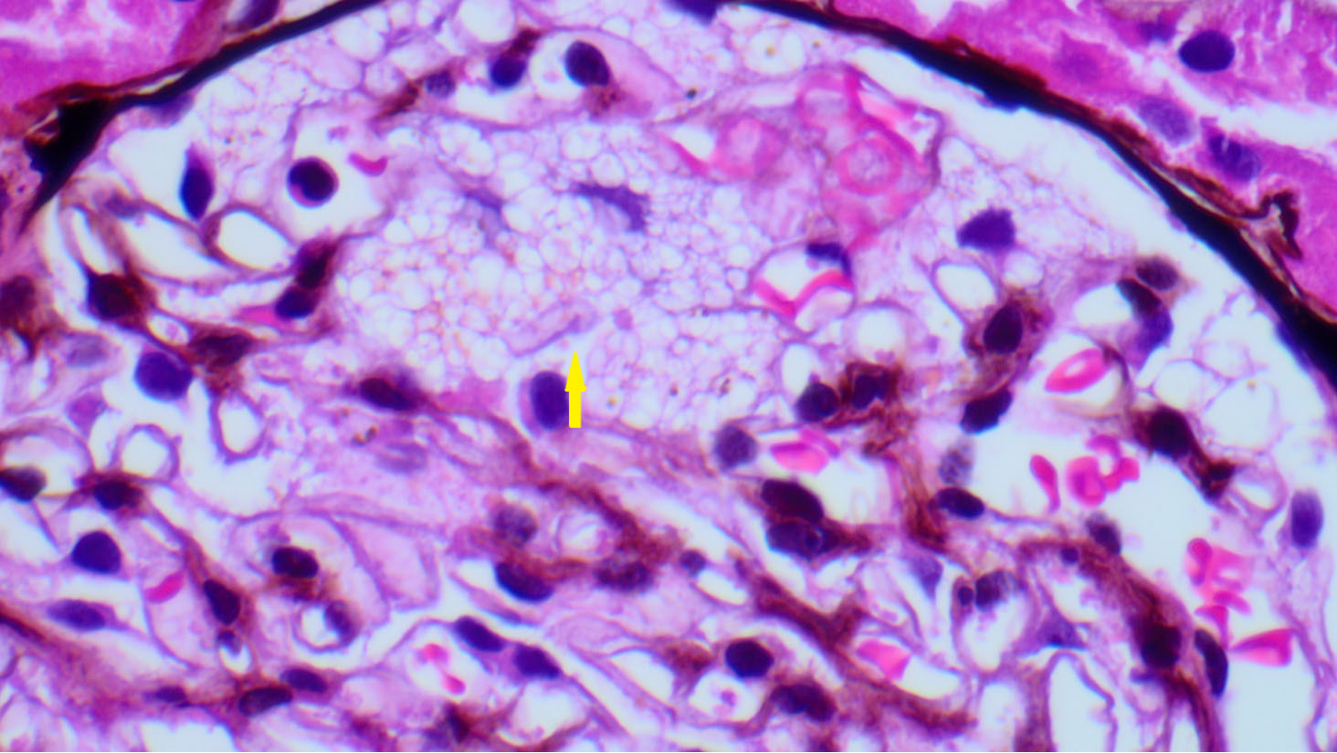
Figure 1. Light microscopy of the kidney biopsy of a female patient. The glomerulus shows swollen podocytes with finely vacuolated cytoplasm (yellow arrow) (H&E; x40).
Figure 1. Light microscopy of the kidney biopsy of a female patient. The glomerulus shows swollen podocytes with finely vacuolated cytoplasm (yellow arrow) (H&E; x40).


All published articles are preserved here permanently:
https://www.portico.org/publishers/oae/


