


fig4
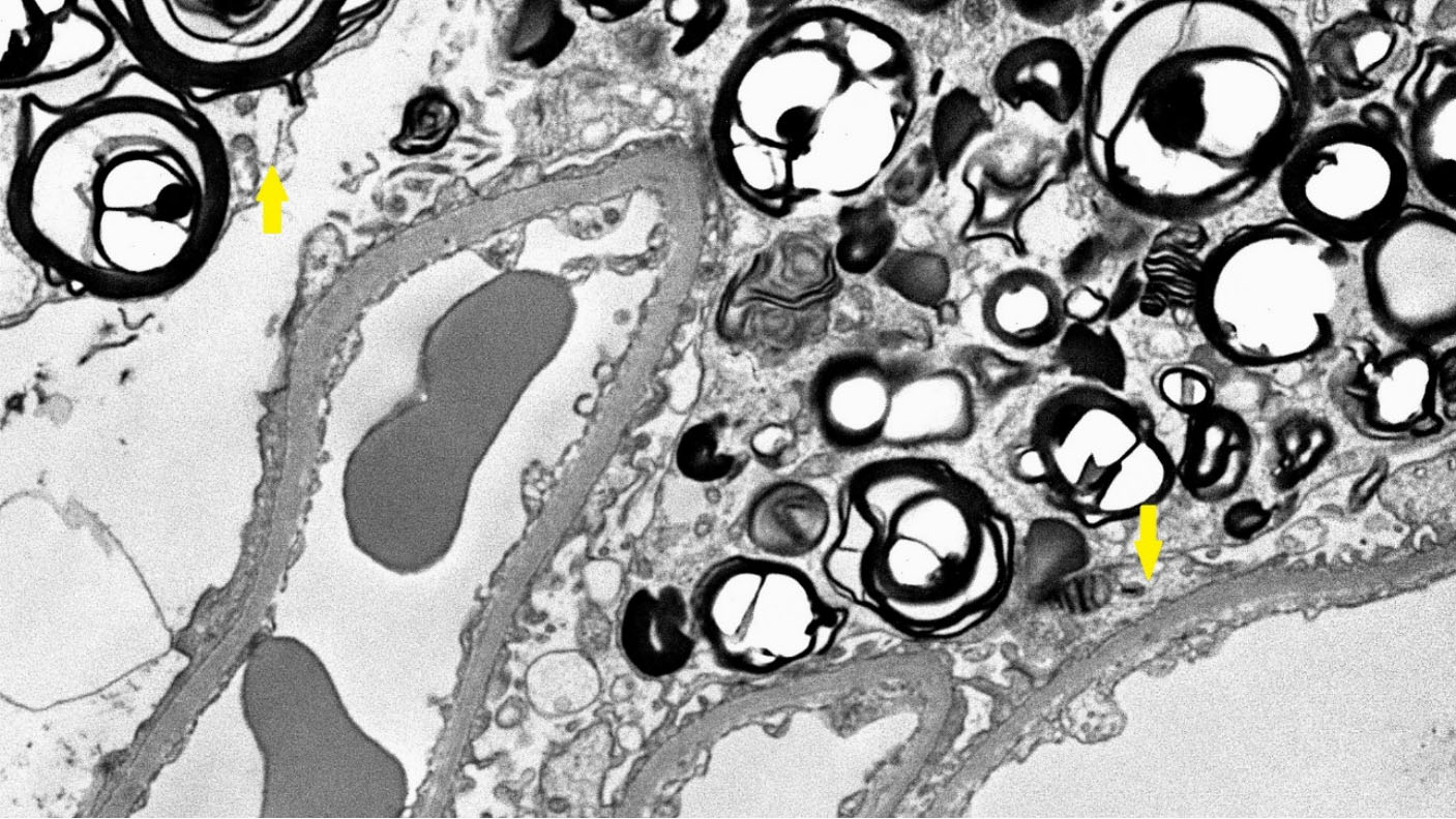
Figure 4. Electron microscopy of the kidney biopsy of a female patient. The glomerulus shows lamellated lipid inclusions (myeloid bodies) in podocytes (yellow arrows).
Figure 4. Electron microscopy of the kidney biopsy of a female patient. The glomerulus shows lamellated lipid inclusions (myeloid bodies) in podocytes (yellow arrows).


All published articles are preserved here permanently:
https://www.portico.org/publishers/oae/


