Nanomedicine-enabled ultrasound immunotherapy: from immunogenic cell death to systemic immune activation
 0
0 Abstract
In recent years, ultrasound-mediated immunotherapy has drawn increasing research interest in cancer immunotherapy due to its non-invasive nature, deep tissue penetration, and spatiotemporal controllability. Ultrasound can induce immunogenic cell death (ICD) through mechanical, cavitation, and sonodynamic effects, thereby promoting the release of tumor antigens and danger-associated molecular patterns (DAMPs) and activating both innate and adaptive immune responses. However, the immune-activating capacity of ultrasound therapy alone remains limited. Advances in nanomedicine have opened new opportunities for ultrasound-mediated immunotherapy. By developing sonosensitizers and piezoelectric, ultrasound-responsive nanoplatforms, it is possible to enhance reactive oxygen species (ROS) production, enable precise drug delivery, and remodel the tumor immune microenvironment, thereby significantly improving antitumor immune efficacy. This article reviews the mechanisms of ultrasound-induced antitumor immunity, nanomedicine-enhanced strategies, regulation of the tumor immune microenvironment, and advances in combination immunotherapy, while discussing the challenges of clinical translation and future directions. Overall, ultrasound immunotherapy is gradually shifting from localized tumor killing to systemic immune activation, offering new insights for precision immunotherapy in solid tumors.
Keywords
INTRODUCTION
Immunotherapy and the challenges facing solid tumor treatment
In recent years, cancer immunotherapy has fundamentally transformed the landscape of traditional cancer treatment by activating the body’s immune system to recognize and eliminate tumor cells. In particular, immune checkpoint blockade (ICB) therapies that target programmed death receptor-1 (PD-1)[1], programmed death ligand-1 (PD-L1)[2], and cytotoxic T-lymphocyte-associated antigen-4 (CTLA-4)[3] have demonstrated significant efficacy in treating various malignancies, including melanoma[4,5], non-small cell lung cancer[6], and renal cell carcinoma[7]. At the same time, chimeric antigen receptor T-cell (CAR-T) therapy has demonstrated unprecedented therapeutic potential in hematologic malignancies[8] [Figure 1]. However, compared with hematologic malignancies, the complex biological characteristics and immunosuppressive microenvironment of solid tumors significantly limit the efficacy of immunotherapy, making it difficult for most patients to achieve durable clinical benefits.
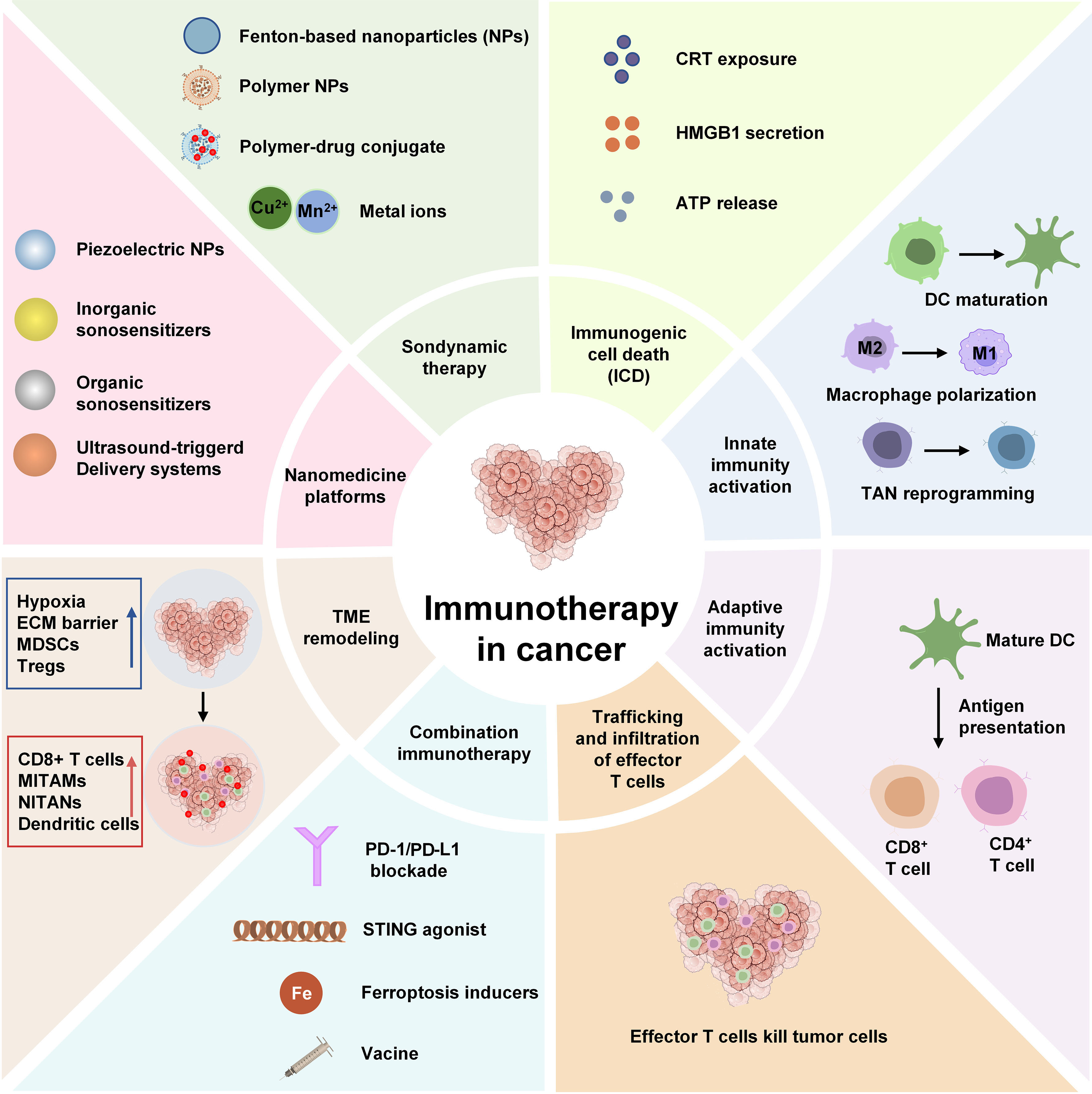
Figure 1. Schematic illustration of nanomedicine-enabled ultrasound immunotherapy for systemic antitumor immune activation. CRT: Calreticulin; DC: dendritic cell; CD8+: cluster of differentiation 8 (positive); CD4+: cluster of differentiation 4 (positive); STING: stimulator of interferon genes; PD-1: programmed death receptor-1; PD-L1: programmed cell death ligand 1; TAN: tumor-associated neutrophil; TME: tumor microenvironment; MDSC: myeloid-derived suppressor cell.
Solid tumors typically exhibit a significantly immunosuppressive tumor microenvironment (TME), characterized by hypoxia[9], an acidic metabolic environment[10], abnormal vascular architecture[11], and a dense extracellular matrix (ECM)[12]. These factors not only hinder the penetration of drugs and immune cells into the deep layers of the tumor but also promote the accumulation of regulatory T cells (Tregs)[13], myeloid-derived suppressor cells (MDSCs)[14], and M2-type tumor-associated macrophages (TAMs)[15], thereby creating a persistent state of immune suppression. Against this backdrop, many solid tumors exhibit a “cold tumor” phenotype characterized by a lack of effector T-cell infiltration, resulting in limited response to ICB therapy. Furthermore, CAR-T cells face challenges in solid tumors, including tumor antigen heterogeneity, low infiltration efficiency, and functional exhaustion[16-18]. Therefore, overcoming the immune barriers in solid tumors and promoting tumor antigen release and immune cell recruitment have become key scientific challenges for improving the efficacy of cancer immunotherapy.
Nanomedicine for overcoming the tumor immune barrier
Advances in nanomedicine have opened up new opportunities for overcoming the immunosuppressive microenvironment of solid tumors. Thanks to their unique size-dependent effects and engineering flexibility, nanocarriers can enhance drug accumulation and retention at the tumor site, enabling the precise delivery of immunomodulators, chemotherapeutic agents, and nucleic acid drugs[19-23]. At the same time, by integrating tumor microenvironment-responsive elements such as pH, reactive oxygen species (ROS), enzymes, and hypoxia, stimulus-responsive nanosystems can enable on-demand drug release and enhance local immune activation[24-28]. In recent years, immuno-nanomedicine has gradually emerged. By deeply integrating nanotechnology with immunotherapy, it enables tumor antigen delivery, immune cell regulation, and tumor microenvironment remodeling[14], demonstrating tremendous potential to improve treatment efficacy and reduce systemic toxicity[29].
However, relying solely on tumor-endogenous stimuli is often limited by tumor heterogeneity and response efficiency, making it difficult to achieve precise spatiotemporal control[30,31]. Therefore, the development of smart nanoplatforms that integrate exogenous stimulation strategies has become a current research focus. Among these, ultrasound - a noninvasive and clinically widely used form of physical stimulation - is attracting increasing attention due to its unique advantages[32,33].
Ultrasound as a noninvasive method of immune modulation
Ultrasound (US) is a mechanical wave with a frequency higher than 20 kHz that enables non-invasive energy delivery to biological tissues[34,35]. Compared to exogenous stimulation methods such as light, magnetic fields, and electric fields, ultrasound has greater tissue penetration capability and can precisely target tissues at the centimeter level or even deeper, making it particularly suitable for the treatment of deep-seated solid tumors[36-38]. Furthermore, focused ultrasound (FUS) technology can precisely concentrate ultrasound energy on the tumor region, providing excellent spatiotemporal controllability and enhancing treatment specificity while minimizing damage to normal tissues[39-42].
At the biological level, ultrasound can induce various physical effects, such as thermal, mechanical, and cavitation effects, thereby enhancing tumor tissue permeability, promoting drug release, and regulating cellular behavior[43-46]. Recent studies have shown that ultrasound is not only a physical therapeutic modality but also an important immunomodulatory tool. Ultrasound-induced cellular stress and tumor cell death can promote the release of tumor-associated antigens and activate the body’s immune system[47-49]. At the same time, ultrasound can improve tumor vascular perfusion, reduce interstitial pressure, and enhance immune cell infiltration, thereby creating favorable conditions for subsequent immunotherapy[50-52].
It is worth noting that ultrasound is already widely used in clinical diagnosis and treatment, including ultrasound imaging, high-intensity focused ultrasound (HIFU) ablation, and ultrasound-assisted drug delivery. This established foundation of clinical applications provides a significant advantage for translating ultrasound-based immunotherapy, giving it greater clinical potential than other emerging stimulation methods.
The rise of ultrasound immunotherapy based on nanomedicine
Advances in nanomedicine have further driven the evolution of ultrasound therapy from simple tumor ablation toward immune activation and immune regulation[53]. By incorporating photosensitizers, piezoelectric materials, immunostimulants, and targeted delivery systems, ultrasound-responsive nanoplatforms can significantly amplify ultrasound-induced ROS production, tumor antigen release, and immunogenic cell death (ICD), thereby initiating the tumor immune cycle[54-57]. At the same time, these nanoplatforms enable the precise delivery of immune checkpoint inhibitors, STING agonists, and cytokines, further enhancing ultrasound-induced antitumor immune responses[58,59].
In recent years, a large body of research has confirmed that ultrasound-responsive nanoplatforms can not only induce local tumor regression but also promote dendritic cell maturation, enhance cluster of differentiation 8 (positive) (CD8+) T-cell infiltration, and establish long-term immune memory, thereby facilitating a transition from local therapy to systemic antitumor immunity[54,60]. This shift in the research paradigm - from “tumor ablation” to “immune activation” and from “local treatment” to “systemic immunity” - is propelling ultrasound immunotherapy to become one of the most promising areas of development in the field of nanomedicine for cancer.
THE BIOLOGICAL BASIS OF ULTRASOUND-INDUCED ANTITUMOR IMMUNITY
Ultrasound-mediated tumor cell death mechanisms
Tumor cell death is the initiating event by which ultrasound immunotherapy triggers the antitumor immune cycle. Unlike conventional therapies, which primarily induce a single apoptotic pathway, ultrasound stimulation can induce multiple forms of cell death, including apoptosis, necrosis, pyroptosis, ferroptosis, and necroptosis[48,61]. Different modes of cell death exhibit varying degrees of immunogenicity, which in turn determine the intensity of subsequent immune activation.
Apoptosis is one of the most common forms of cell death in ultrasound therapy. Ultrasound activates photosensitizers, generating large amounts of ROS, which cause mitochondrial damage, the release of cytochrome C, and the activation of the caspase cascade, ultimately leading to programmed cell death[62-64]. However, classical apoptosis is typically accompanied by the complete encapsulation and clearance of cellular contents, resulting in a mild inflammatory response. Therefore, its immunogenicity is relatively limited.
In contrast, ultrasound-induced necrosis can disrupt cell membrane integrity, resulting in the release of large amounts of intracellular components into the extracellular space. The released nucleic acids, proteins, and metabolic byproducts can be recognized by the body as danger signals, thereby inducing a local inflammatory response and the recruitment of immune cells[65]. Although necrosis is highly immunogenic, its uncontrolled nature may damage normal tissues.
Recent studies have shown that inflammatory programmed cell death processes such as pyroptosis, ferroptosis, and necroptosis play an increasingly important role in ultrasound immunotherapy. Pyroptosis is a form of programmed inflammatory cell death mediated by Gasdermin proteins. Ultrasound-induced ROS can activate the NOD-like receptor family pyrin domain containing 3 (NLRP3) inflammasome, promoting Caspase-1-mediated cleavage of Gasdermin D, which creates pores in the cell membrane and ultimately leads to the massive release of inflammatory factors such as interleukin-1 beta (IL-1β), interleukin-18 (IL-18), and ATP[66-68]. Because pyroptosis is accompanied by a strong inflammatory response, it is considered a crucial bridge linking tumor cell death to immune activation.
Ferroptosis is a form of cell death that depends on iron ions and lipid peroxidation[69]. Ultrasound-induced ROS promotes the accumulation of lipid peroxides and inhibits glutathione peroxidase 4 (GPX4) activity, thereby triggering ferroptosis[70,71]. Studies have shown that the oxidized lipids and danger signals released during ferroptosis promote dendritic cell maturation and CD8+ T-cell infiltration, making it a subject of widespread interest in the field of tumor immunotherapy.
Necroptosis is a form of programmed cell death regulated by the RIPK1-RIPK3-MLKL signaling axis [72]. Compared with apoptosis, necroptosis releases more danger-associated molecular patterns (DAMPs) and induces a more intense immune response[73]. A growing body of evidence suggests that ultrasound-induced necroptosis may be one of the key mechanisms for activating subsequent antitumor immunity.
Different types of cell death exhibit significant differences in immunogenicity, and their ability to induce antitumor immune responses varies. Recent studies have shown that programmed cell death pathways such as pyroptosis, necroptosis, and ferroptosis typically exhibit stronger immunostimulatory effects, while ultrasound-induced ICD is considered a crucial bridge linking localized tumor killing to systemic immune activation. A summary of the main characteristics and immunological effects of different cell death modes is presented in Table 1.
Comparison of immunogenicity among different ultrasound-induced tumor cell death pathways
| Modes of cell death | Molecular mechanism | Biomarkers | Immunogenicity | Antitumor immune effects | Ref. |
| Apoptosis | Caspase-3/8/9 activation | Cleaved caspase-3, Annexin V | Low-Moderate | Limited antigen release and weak immune stimulation | [63] |
| Necrosis | Plasma membrane rupture | LDH release | Moderate | Induces inflammation but may cause nonspecific tissue damage | [65] |
| Necroptosis | RIPK1/RIPK3/MLKL pathway | p-MLKL, RIPK3 | High | Promotes dendritic cell activation and T-cell priming | [72] |
| Pyroptosis | Caspase-1/GSDMD signaling | IL-1β, IL-18, GSDMD-N | High | Strong activation of innate immune responses | [74] |
| Ferroptosis | Iron-dependent lipid peroxidation | GPX4, ACSL4, MDA | High | Enhances DAMP release and T-cell recruitment | [70,75] |
| Immunogenic Cell Death (ICD) | ER stress, DAMP emission (CRT exposure, ATP release, HMGB1 secretion) | CRT, ATP, HMGB1, HSP70/90 | Very High | As immunogenic outcome shared by several regulated cell-death modalities | [76] |
Immunogenic cell death: the starting point of immune activation
Among the various forms of cell death, ICD is considered a key bridge linking localized tumor killing to systemic immune activation. Unlike conventional forms of cell death, ICD not only eliminates tumor cells but also transforms the dead cells into natural “tumor vaccines,” thereby activating the body’s immune system.
The most important feature of ICD is the exposure and release of DAMPs. Among these, the exposure of calreticulin (CRT) on the cell surface is an early marker of ICD. As a typical “Eat-me signal,” CRT promotes the recognition and phagocytosis of dead tumor cells by dendritic cells and macrophages, thereby enhancing antigen uptake efficiency[77]. At the same time, the release of extracellular ATP constitutes a “Find-me signal,” which recruits dendritic cells, macrophages, and neutrophils to the tumor site via purinergic receptor signaling pathways, thereby promoting the initiation of local immune responses[78]. High-mobility group box protein 1 (HMGB1) is an important DAMP molecule released during the later stages of ICD[79]. Upon binding to TLR4 on the surface of dendritic cells, HMGB1 promotes antigen processing and cross-presentation, thereby enhancing the efficiency of antigen-specific T-cell activation[80]. Furthermore, the exposure of heat shock proteins heat shock protein 70(HSP70) and HSP90 also enhances tumor antigen presentation and immune recognition[81].
In recent years, a large body of research has shown that ultrasound-responsive nanoplatforms can significantly enhance ICD through ROS bursts, endoplasmic reticulum stress, and mitochondrial damage. For example, the large amounts of ROS generated by sonodynamic therapy can induce an endoplasmic reticulum stress response, promoting CRT translocation and ATP release. Piezoelectric nanomaterials, in turn, can further amplify DAMPs release through sustained oxidative stress. Therefore, ICD is widely recognized as the core event that initiates the antitumor immune cycle in ultrasound immunotherapy.
Activation of the innate immune system
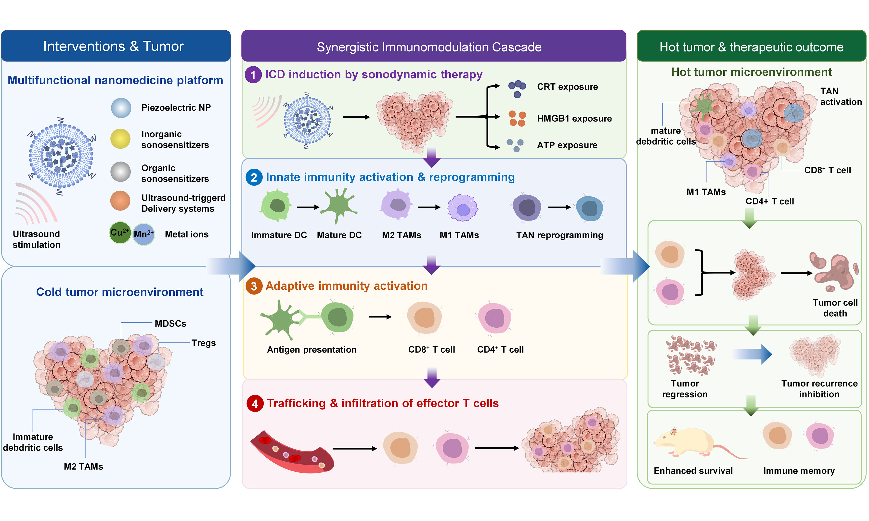
DAMPs and tumor-associated antigens produced by ICDs further activate the body’s innate immune system, laying the foundation for subsequent adaptive immune responses. Dendritic cells (DCs) serve as a central hub linking the innate and adaptive immune systems. Upon stimulation by DAMPs, immature DCs gradually mature and upregulate the expression of CD80, CD86, and major histocompatibility complex (MHC) molecules, thereby enhancing their ability to process and present tumor antigens[82,83]. However, DC metabolic dysregulation often leads to the premature transport of antigens to lysosomes for degradation, resulting in reduced antigen presentation efficiency, which severely limits the quality and intensity of T-cell activation. To address this bottleneck, Chen et al. developed an ultrasound-activatable in situ vaccine based on a thylakoid/platelet hybrid membrane-camouflaged nanovesicle (TPLHZ) that co-delivers the DNA methyltransferase inhibitor zebularine and the sonosensitizer hematoporphyrin monomethyl ether[83]. Beyond boosting ROS-mediated ICD and antigen release, zebularine effectively reverses tumor DNA hypermethylation, upregulating MHC-I expression on tumor cells to enhance antigen self-presentation, while the ICD-triggered DAMPs synergistically promote DC maturation and cross-presentation. This dual-regulation strategy significantly amplifies T-cell activation, elicits potent cytotoxic CD8+ T-cell responses, establishes long-term immunological memory (characterized by increased central and effector memory T-cell populations), and demonstrates robust synergistic efficacy with immune checkpoint blockade, offering a promising approach to overcome anti-PD-1 resistance in solid tumors [Figure 2].
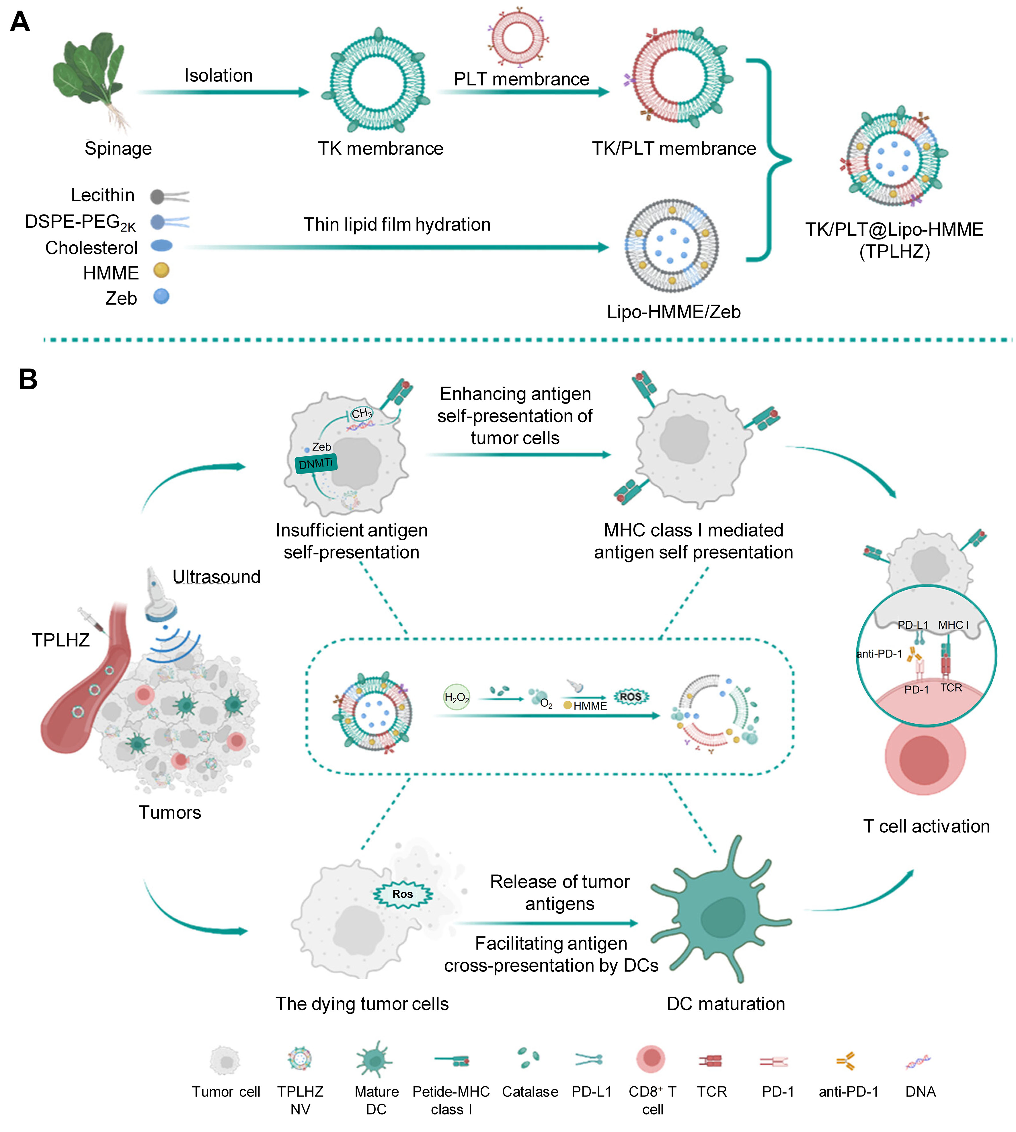
Figure 2. Schematic illustration of the ultrasound-activatable in situ vaccine strategy for enhanced antigen self-and cross-presentation to overcome resistance. (A) Preparation process of TPLHZ; (B) Under ultrasound irradiation, TPLHZ is anticipated to inhibit tumor DNA hypermethylation and promote MHC-I-mediated antigen self-presentation. Meanwhile, excessive ROS production can lead to the in situ release of tumor antigens from dying cells and facilitate the cross-presentation of tumor antigens by DCs. Enhanced antigen self- and cross-presentation potentiates tumor immunotherapy. Reproduced with permission[83]. Copyright 2024, ACS Nano. PLT: Platelet; TK: thylakoid; HMME: hematoporphyrin monomethyl ether; TCR: T-cell receptor; PD-1: programmed death-1; PD-L1: programmed death-ligand 1; ROS: reactive oxygen species; MHC: major histocompatibility complex; DC: dendritic cell; TPLHZ: TK/PLT@Lipo-HMME/Zeb; DNMTi: DNA methyltransferase inhibitor; DSPE: 1,2-Distearoyl-sn-glycero-3-phosphoethanolamine; PEG: poly(ethylene glycol); Zeb: zebularine; NV: nanovesicle.
Tumor-associated macrophages (TAMs) are among the most abundant immune cells in the tumor microenvironment. In most solid tumors, TAMs primarily exhibit a pro-tumor M2 phenotype. Studies have shown that ultrasound-induced ROS and inflammatory factors can promote the conversion of M2 macrophages to M1 macrophages[59]. M1 macrophages secrete pro-inflammatory cytokines such as TNF-α, IL-12, and IFN-β, which enhance antigen presentation and promote T-cell activation.
In recent years, the role of neutrophils in ultrasound-mediated immunotherapy has gradually garnered attention [Figure 3]. Tumor-associated neutrophils also exhibit two functional states: N1 and N2[84]. The ultrasound-induced inflammatory microenvironment can promote the conversion of N2-type neutrophils to N1-type neutrophils[85]. N1-type neutrophils can release ROS, TNF-α, and chemokines, and promote the recruitment of CD8+ T cells, thereby enhancing the antitumor immune response[86,87].
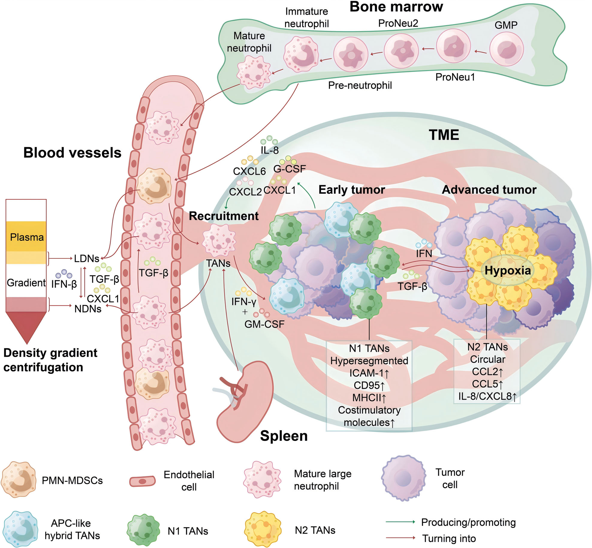
Figure 3. Two main phenotypes of tumor-associated neutrophils within the tumor microenvironment. Reproduced with permission[84]. Copyright 2025, Signal Transduction and Targeted Therapy. TME: Tumor microenvironment; PMN-MDSC: polymorphonuclear myeloid-derived suppressor cell; NDN: normal-density neutrophil; LDN: low-density neutrophil; GMP: granulocyte-macrophage progenitor; IL-8: interleukin-8; TGF: transforming growth factor; IFN: interferon; CXCL: CXC-chemokine ligand; TAN: tumor-associated neutrophil; APC: antigen-presenting cell; GM-CSF: granulocyte-macrophage colony-stimulating factor; ICAM-1: intercellular cell adhesion molecule-1; CD95: cluster of differentiation 95; MHCII: major histocompatibility complex class II; CCL: chemokine (C-C motif) ligand; G-CSF: granulocyte colony-stimulating factor.
In addition, natural killer (NK) cells are also involved in the early immune response induced by ultrasound. Increased expression of stress ligands on the surface of damaged tumor cells promotes NK cell activation and enhances their ability to release perforin and granzyme, thereby further eliminating tumor cells[88,89].
Establishment of the adaptive immune response
The adaptive immune response is a key factor determining the long-term efficacy of ultrasound-mediated immunotherapy. Mature dendritic cells present captured tumor antigens to naive T cells, thereby initiating an antigen-specific immune response.
CD8+ cytotoxic T lymphocytes are the primary effector cells of antitumor immunity. Under MHC-I molecule-mediated antigen presentation, CD8+ T cells undergo clonal expansion and migrate to tumor tissue, where they directly kill tumor cells by releasing perforin and granzyme[90,91]. Numerous studies have confirmed that one of the key indicators of successful ultrasound immunotherapy is a significant increase in CD8+ T-cell infiltration within tumor tissue. CD4+ helper T cells support the expansion and functional maintenance of CD8+ T cells by secreting cytokines such as IL-2, IFN-γ, and TNF-α, while also coordinating the joint participation of B cells and innate immune cells in the antitumor response[92-94].
Notably, the massive release of tumor antigens induced by ultrasound can also increase T-cell receptor (TCR) repertoire diversity, enabling the body to recognize more tumor neoantigens and thereby reducing the risk of tumor immune evasion[95]. Furthermore, some effector T cells can further differentiate into memory T cells, including central memory T cells and effector memory T cells[96,97]. These cells persist in the body for extended periods and can rapidly mount an immune response upon tumor recurrence, ensuring long-term antitumor immune surveillance.
From local immune activation to systemic antitumor immunity
The most compelling feature of ultrasound immunotherapy lies in its ability to transform localized treatment into a systemic antitumor immune response. Although the ultrasound effect is confined to the local lesion, the immune response it induces can spread throughout the body via the circulatory system.
The abscopal effect is a classic manifestation of systemic immune activation. Following ultrasound treatment of the primary tumor, large numbers of activated tumor-specific T cells enter the circulatory system and migrate to distant metastatic sites, thereby inhibiting the growth of tumors that have not been directly treated[98]. A growing body of research indicates that combining ultrasound with immune checkpoint blockade therapy can significantly enhance the abscopal effect and improve control of metastatic tumors.
Systemic immune activation can also effectively eliminate circulating tumor cells (CTCs) and micrometastases, thereby reducing the risk of tumor metastasis. At the same time, the formation of memory T cells endows the body with long-term immune surveillance capabilities, enabling it to rapidly mount an immune response upon tumor recurrence and prevent tumor regrowth[99-101].
Therefore, the ultimate goal of ultrasound-mediated immunotherapy is no longer limited to localized tumor ablation but rather involves transitioning from localized treatment to systemic antitumor immunity by inducing ICD, activating innate and adaptive immunity, and establishing immune memory. This process constitutes a complete biological chain of logic “from immunogenic cell death to systemic immune activation” and provides an important theoretical foundation for the design of future ultrasound-mediated immunotherapy strategies empowered by nanomedicine.
STRATEGIES FOR ENHANCING ULTRASOUND-ASSISTED IMMUNOTHERAPY USING NANOMEDICINE
Although ultrasound can induce tumor cell death and activate antitumor immunity through mechanical, cavitation, and thermal effects, its use as a standalone therapy still faces challenges such as limited ROS production efficiency, insufficient immune activation, and immune suppression in the tumor microenvironment[104,105]. Advances in nanomedicine offer new approaches to addressing these bottlenecks. By developing functional nanoplatforms with acoustic responsiveness, it is possible not only to significantly improve the efficiency of ultrasound energy utilization but also to achieve precise drug delivery, regulation of the tumor microenvironment, and immune enhancement, thereby transforming ultrasound-induced localized tumor damage into a sustained and robust systemic antitumor immune response.
With the continuous development of ultrasound-responsive nanoplatforms, researchers have constructed a variety of nanosystems, including sound-sensitive platforms, piezoelectric platforms, ultrasound-controlled drug delivery systems, and biomimetic platforms[106-108]. These platforms each offer distinct advantages in ROS generation, drug delivery, immune regulation, and tumor targeting, and collectively drive the evolution of ultrasound immunotherapy from localized tumor destruction to systemic immune activation. A comparison of key parameters for different ultrasound-responsive nanomaterials is presented in Table 2.
Representative nanomedicine strategies for enhancing ultrasound immunotherapy and their immune-regulatory functions
| Nanoplatform category | Representative materials | Ultrasound- responsive mechanism | Immune Effects | Advantages | Ref. |
| Organic sonosensitizers | Ce6, HMME, porphyrins | Sonodynamic ROS generation | ICD induction, DCs maturation | High biocompatibility | [21] |
| Inorganic sonosensitizers | TiO2, MnOx, Black Phosphorus | Sonocatalytic ROS production | Enhanced CD8+ T-cell infiltration | High ROS generation efficiency | [101] |
| Metal-organic frameworks (MOFs) | Porphyrin-based bimetallic MOFs (Mn-Fe TCPP), D-A structured MOFs (BTP) | Sonocatalytic ROS generation via ordered confinement or D-A architecture | ICD induction, DC maturation, ferroptosis, cGAS-STING activation | High porosity, structural tunability, minimized π-π aggregation, dual-metal synergistic effects | [102] |
| MXenes | MXene@MnO2-TPP Schottky heterojunctions (Ti3C2Tx@MnO2) | Schottky heterojunction-enhanced electron-hole separation under ultrasound | Ferroptosis induction, mtDNA release, cGAS-STING activation, systemic antitumor immunity | Narrow bandgap, high charge separation efficiency, mitochondria-targeting capability | [103] |
| Piezoelectric nanoplatforms | BaTiO3, ZnO, BiOCl, KNN | Piezoelectric polarization-induced electron-hole separation | ICD induction, M1 polarization, DC activation | No exogenous sensitizer required | [107] |
| Ultrasound-triggered drug delivery systems | Liposomes, hydrogels, polymeric nanoparticles | Acoustic cavitation-triggered payload release | Enhanced immunomodulator delivery | Spatiotemporal controllability | [108] |
| Biomimetic nanoplatforms | Tumor cell membrane, DC membrane, macrophage membrane, neutrophil membrane | Biomimetic targeting and immune evasion | Improved antigen presentation and tumor homing | Prolonged circulation and targeting capability | [99] |
Organic sonosensitizers offer superior biocompatibility but suffer from poor stability, whereas inorganic counterparts exhibit higher ROS efficiency yet raise toxicity concerns. Piezoelectric platforms eliminate the need for photosensitizers but vary substantially in ROS yield across materials due to differences in piezoelectric properties, and their performance depends on poorly standardized ultrasound parameters. Ultrasound-triggered drug carriers enable spatiotemporal release, yet liposomes risk premature leakage, polymeric nanoparticles suffer from attenuated responsiveness in deep tumors, and hydrogels require invasive administration. These platform-specific trade-offs underscore the need for disease-specific selection and standardized evaluation criteria, rather than pursuing a single optimal platform.
REMODELING OF THE TUMOR IMMUNE MICROENVIRONMENT
The tumor immune microenvironment (TIME) comprises a diverse array of components, including tumor cells, immune cells, stromal cells such as cancer-associated fibroblasts (CAFs), vascular networks, and the extracellular matrix (ECM). Its dynamic changes directly determine tumor progression and the efficacy of immunotherapy. In most solid tumors, the TIME typically exhibits a highly immunosuppressive state, characterized by the accumulation of pro-tumor immune cells, insufficient infiltration of effector T cells[109], abnormal vascular structure[110], and excessive ECM deposition[111]. Notably, CAFs mediate significant immune evasion and physical barrier effects through the secretion of cytokines and the construction of a dense ECM barrier. These factors not only limit drug delivery efficiency but also weaken the body’s antitumor immune response. Recent studies have shown that ultrasound-responsive nanotechnology platforms can not only induce tumor cell death but also reprogram the tumor microenvironment by regulating immune cell function, improving vascular structure, and remodeling the stromal barrier, thereby creating favorable conditions for establishing a systemic antitumor immune response.
The remodeling of the tumor immune microenvironment does not depend on a single type of immune cell but involves dynamic, coordinated regulation among multiple cell populations, including tumor-associated macrophages, tumor-associated neutrophils, myeloid suppressor cells, dendritic cells, effector T cells, and CAFs[112]. The ultrasound-responsive nanoplatform facilitates the transition from an immunosuppressive microenvironment to an immune-activating state by simultaneously acting on these key immune components. The relevant regulatory mechanisms are summarized in Table 3.
Regulation of major immune cell populations during ultrasound-mediated tumor microenvironment remodeling
| Immune component | Pro-tumor phenotype | Anti-tumor phenotype | Ultrasound-mediated regulation | Immunological outcome | Ref. |
| Tumor-associated macrophages (TAMs) | M2 | M1 | ROS generation, inflammatory cytokine production | enhanced antigen presentation and T-cell activation | [21] |
| Tumor-associated neutrophils (TANs) | N2 | N1 | ROS signaling and TGF-β inhibition | increased tumor cytotoxicity and immune recruitment | [84] |
| Myeloid-derived suppressor cells (MDSCs) | immunosuppressive | reduced abundance | STAT3/TGF-β pathway inhibition | relief of immunosuppression | [14] |
| Dendritic cells (DCs) | immature | mature | DAMP-mediated activation | enhanced antigen uptake and cross-presentation | [83] |
| Natural killer cells (NK Cells) | dysfunctional | activated | stress ligand upregulation and cytokine stimulation | increased innate immune cytotoxicity | [89] |
| Tumor vasculature | abnormal | normalized | improved perfusion and reduced hypoxia | enhanced immune-cell infiltration | [110] |
| Extracellular matrix (ECM) | dense barrier | remodeled matrix | mechanical disruption and enzyme-assisted degradation | improved T-cell penetration | [111] |
| Cancer-associated fibroblasts (CAFs) | Activated CAFs (secrete TGF-β, IL-6, CXCL12, collagen) | inactivated/ depleted CAFs | ultrasound-induced mechanical forces, CAF-targeted sonodynamic therapy (SDT), ECM softening and stromal depletion | reduced ECM density, enhanced T-cell infiltration, improved drug/immune cell penetration, relief of immunosuppression | [112] |
The immunomodulatory effects of these nanoplatforms involve coordinated regulation of multiple immune populations, yet their relative contributions vary across tumor models. While TAM polarization and DC maturation are consistently observed, neutrophil phenotype switching appears more microenvironment-dependent. This heterogeneity underscores the need for comprehensive immune profiling to guide rational combination strategies.
COMBINED TREATMENT STRATEGIES FOR SYSTEMIC IMMUNE ACTIVATION
Although ultrasound immunotherapy can activate antitumor immune responses by inducing ICD, monotherapy often struggles to overcome the complex immunosuppressive microenvironment and maintain long-term, effective immune surveillance. Recent studies have shown that ultrasound therapy is better suited to serve as a “trigger” for the Cancer-Immunity Cycle. Crucially, the efficacy of this trigger in synergizing with immune checkpoint blockade (ICB) depends on the precise calibration of ultrasound parameters and the resulting ICD intensity. Optimal outcomes are typically achieved within a defined “therapeutic window,” such as acoustic pressures of 1.0-1.5 MPa and duty cycles of ~ 50%[113]. Within this range, ultrasound induces a moderate-to-high level of ICD - characterized by robust CRT exposure, HMGB1 translocation, and ATP secretion - which effectively converts “cold” tumors into “hot” ones by enhancing dendritic cell maturation and T-cell priming. Conversely, overtreatment leading to massive necrosis may exacerbate immunosuppression via HIF-1α stabilization and adenosine accumulation, thereby compromising ICB efficacy. Thus, combining ultrasound with other immunotherapy strategies not only amplifies the immune response but also establishes persistent systemic antitumor immunity[114]. Consequently, developing combination therapy regimens centered on strategies such as immune checkpoint blockade, STING activation, ferroptosis, adoptive cell therapy, and tumor vaccines has become the most active area of research in the field of ultrasound immunotherapy.
Ultrasound-guided immunotherapy combined with immune checkpoint inhibition
Ultrasound-induced SDT can lead to massive tumor cell death, accompanied by ICD hallmarks such as CRT exposure, ATP release, and HMGB1 secretion. When tumor-associated antigens and DAMPs are taken up by dendritic cells, they promote dendritic cell maturation and enhance their ability to cross-present antigens, ultimately inducing the expansion of tumor-specific CD8+ T cells. However, once activated T cells enter tumor tissue, they are often suppressed by immune checkpoint signaling, such as PD-1/PD-L1 and CTLA-4.
PD-1 is a negative regulatory receptor expressed on the surface of activated T cells. Upon binding to PD-L1 on the surface of tumor cells, it inhibits TCR signaling, reduces the release of cytotoxic molecules (granzyme B and perforin), and induces T-cell exhaustion. At the same time, the ultrasound-induced inflammatory response and IFN-γ release may, conversely, further promote PD-L1 expression, leading to adaptive immune tolerance.
The sound-activated semiconductor polymer nano-feedback system (SPNSA) developed by Yu et al. takes advantage of this mechanism: Upon ultrasonic activation, the H2O2 generated by SPNs not only mediates ICD induced by sonodynamic therapy but also cleaves the linker to release the loaded aPD-L1, precisely blocking the upregulated PD-L1 pathway[115]. This acts synergistically with antigen release induced by SDT, achieving simultaneous suppression of primary and distant tumors in a bilateral 4T1 tumor model[115]. In addition to PD-L1, the adenosine/A2AR pathway is also a key immunosuppressive feedback mechanism following ICD. By using SPNSA to simultaneously release the A2AR antagonist SCH58261, the research team achieved dual-pathway blockade, which further amplified the antitumor immune response [Figure 4].
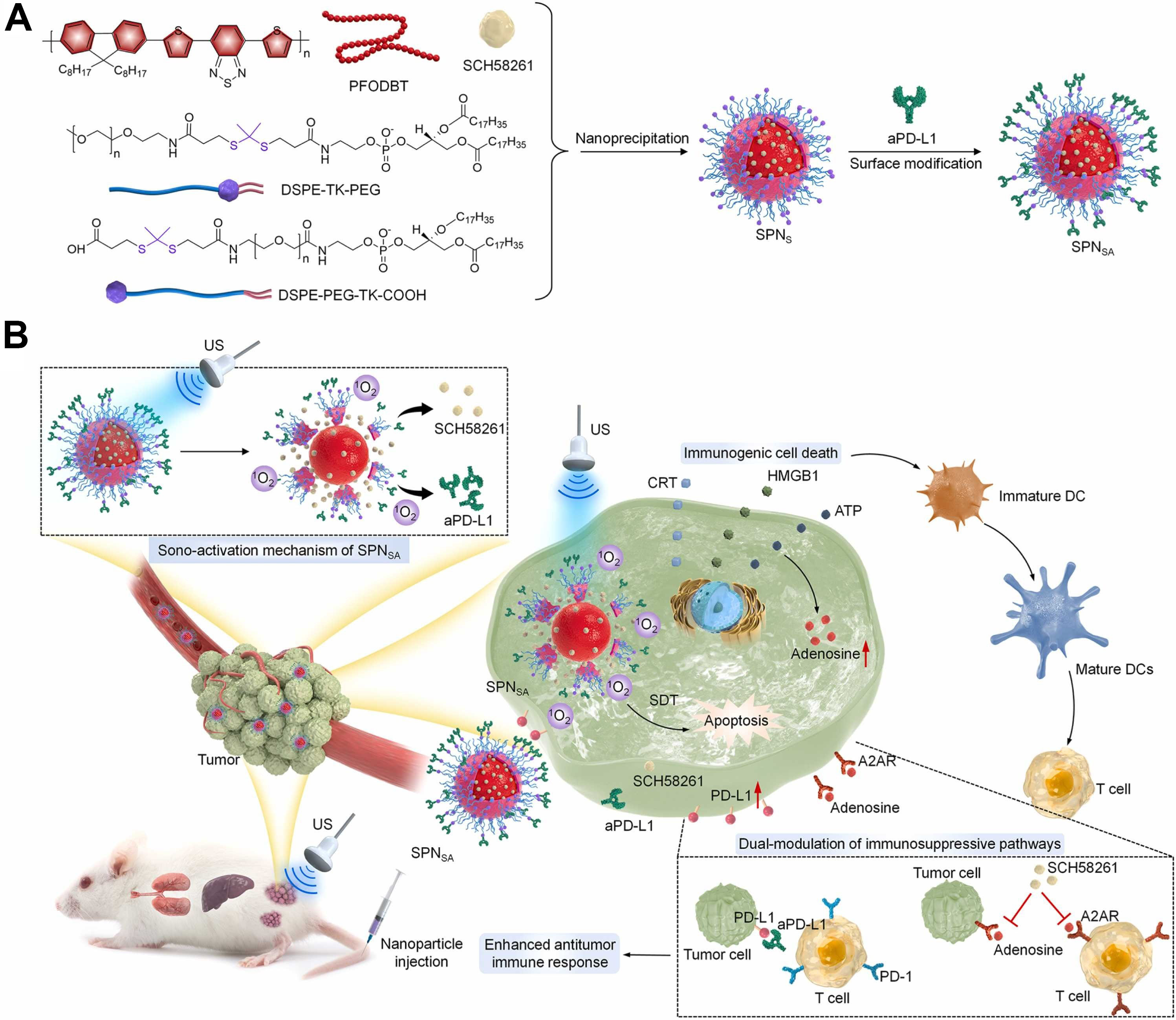
Figure 4. Development of sono-activatable nanofeedbacks (SPNSA) for precision dual-modulation of immunosuppressive pathways and SDT-combinational immune therapy. (A) Construction of SPNSA via nanoprecipitation and surface modification; (B) Schematic illustration of the mechanisms of sono-activatable SPNSA for ICD induction, dual-modulation of immunosuppressive pathways, and SDT-combinational immunotherapy. Reproduced with permission[115]. Copyright 2023, Nano Today. SDT: Sonodynamic therapy; aPD-L1: anti-programmed death-ligand 1 antibody; PD-1: programmed cell death protein 1; PD-L1: programmed death-ligand 1; DC: dendritic cell; ICD: immunogenic cell death; PFODBT: poly[2,7-(9,9-di-octyl-fluorene)-alt-4,7-bis(thiophen-2-yl)benzo-2,1,3-thiadiazole]; DSPE-TK-PEG: 1,2-Distearoyl-sn-glycero-3-phosphoethanolamine-thioketal-poly(ethylene glycol); DSPE-PEG-TK-COOH: 1,2-Distearoyl-sn-glycero-3-phosphoethanolamine-poly(ethylene glycol)-thioketal-carboxylic acid; US: ultrasound.
CTLA-4 primarily acts during the T-cell activation phase by competitively binding to CD80/CD86 molecules on the surface of dendritic cells, thereby inhibiting the formation of co-stimulatory signals and limiting initial T-cell activation. The massive release of tumor antigens induced by ultrasound provides the foundation for T-cell activation, while CTLA-4 blockade further enhances T-cell clonal expansion[116]. In the future, therapeutic strategies combining ultrasound immunotherapy with CTLA-4 blockade are expected to be developed. By enhancing T-cell priming and clonal expansion, these strategies can effectively reverse the tumor-suppressive immune microenvironment, thereby further enhancing the level of antitumor immune response[117]. The combination of these two approaches not only enhances local tumor killing but also promotes the formation of memory T cells, thereby establishing long-term immune surveillance.
Ultrasound-assisted immunotherapy combined with STING activation
The cGAS-STING pathway is a key signaling axis through which the body recognizes cytoplasmic DNA and initiates innate immune responses, playing a central role in linking innate and adaptive immunity. Ultrasound-induced ROS-mediated damage and cell death can lead to the release of nuclear and mitochondrial DNA from tumor cells into the cytoplasm[118]. Upon recognition of cytoplasmic DNA by cyclic GMP-AMP synthase (cGAS), cGAS catalyzes the production of the second messenger cyclic guanosine monophosphate–adenosine monophosphate (cGAMP), which subsequently activates the STING protein on the endoplasmic reticulum. Activated STING recruits TANK-binding kinase 1 (TBK1) and promotes the phosphorylation of IRF3, thereby inducing the production of large amounts of type I interferons (IFN-α/β). Type I interferons not only promote dendritic cell maturation but also enhance their ability to cross-present antigens and induce the expression of chemokines such as CXCL9 and CXCL10, thereby facilitating the recruitment of CD8+ T cells to tumor tissues. Consequently, the STING pathway is considered a key switch for initiating the immune response in “cold tumors”.
However, because some tumor cells exhibit STING signaling defects or insufficient accumulation of cytoplasmic DNA, relying solely on ultrasound-induced endogenous DNA release often fails to fully activate the cGAS-STING pathway. To address this issue, Lei et al. developed a mitochondria-targeted sonodynamic nanoplatform, TPP@CoTCPP, which enhances ultrasound-induced ROS production through TPP-mediated mitochondrial targeting[119]. Abundant ROS induce mitochondrial damage and mitochondrial permeability transition pore (mPTP) opening, promoting the release of mtDNA into the cytoplasm and thereby activating the cGAS-STING-TBK1-IRF3 signaling axis [Figure 5]. Further studies have shown that this strategy not only significantly enhances the expression of STING-related signaling molecules but also promotes the transition of macrophages to a pro-inflammatory phenotype. Together with sonodynamic therapy-induced immunogenic cell death, it amplifies the antitumor immune response, providing a new design approach for utilizing ultrasound to activate the innate immune system.
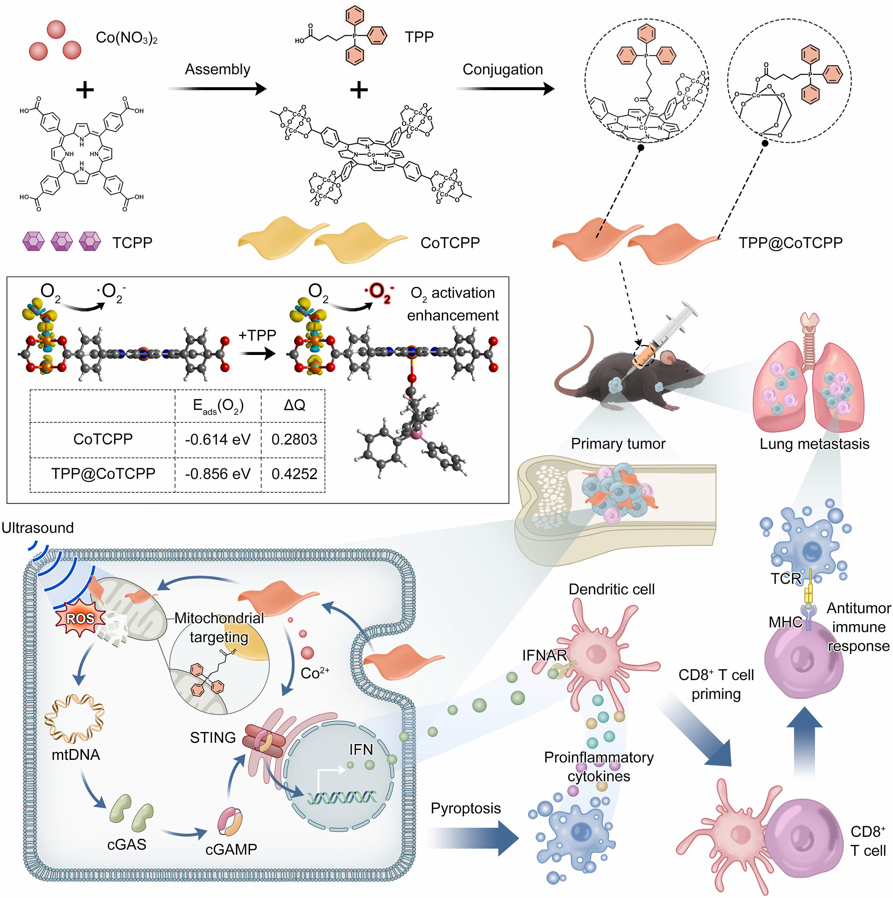
Figure 5. Graphic illustration of the sonodynamic-ionicimmunotherapy mechanism. Reproduced with permission[119]. Copyright 2023, Nano Today. Abbreviations: TCPP: tetra(4-carboxyphenyl)porphyrin; TPP: tetra(pentafluorophenyl)porphyrin; STING: stimulator of interferon genes; cGAMP: cyclic guanosine monophosphate–adenosine monophosphate; IFN: interferon; IFNAR: interferon-alpha/beta receptor; CD8+: cluster of differentiation 8 (positive); ROS: reactive oxygen species; mtDNA: mitochondrial DNA; cGAS: cyclic GMP-AMP synthase; TCR: T cell receptor; MHC: major histocompatibility complex.
Ultrasound immunotherapy combined with ferroptosis therapy
Ferroptosis is a form of programmed cell death driven by iron-dependent lipid peroxidation, and its progression exhibits a high degree of synergy with ultrasound-induced ROS production. The large amount of ROS generated during SDT continuously attacks the polyunsaturated fatty acids in the cell membrane, leading to the continuous accumulation of lipid peroxides. When GPX4 activity is inhibited, cells are unable to clear lipid peroxides in a timely manner, ultimately inducing ferroptosis[120]. At the same time, iron ions continuously generate hydroxyl radicals via the Fenton reaction, further amplifying oxidative damage.
Compared with conventional apoptosis, ferroptosis exhibits greater immunogenicity; ferroptotic cells release signaling molecules such as oxidized phospholipids, HMGB1, and ATP, thereby promoting dendritic cell maturation and antigen uptake. Furthermore, activated CD8+ T cells release IFN-γ, which downregulates solute carrier family 7 member 11 (SLC7A11) expression in tumor cells, further enhancing sensitivity to ferroptosis[121]. The continuous inertial cavitation strategy developed by Yin et al. induces the generation of large amounts of ROS via ultrasound, which synergizes with ferroptosis inducers to significantly enhance lipid peroxidation levels and ICD effects, thereby achieving highly effective tumor suppression and immune activation in a breast cancer model[122]. Similarly, the dual-programmed sonodynamic-ferroptosis nano-PROTAC platform (SPNFeP) developed by Wang et al. utilizes the synergistic amplification of oxidative stress through ultrasound-triggered singlet oxygen generation and hydroxyl radicals produced by the Fenton reaction[123]. This significantly promotes lipid peroxidation and GPX4 downregulation, inducing intense ferroptosis and ICD responses, as evidenced by increased CRT exposure, ATP release, and HMGB1 secretion; This further enhances dendritic cell maturation and CD8+ T-cell infiltration while reducing MDSC accumulation, thereby achieving effective immune activation and complete suppression of deep-seated tumors [Figure 6].
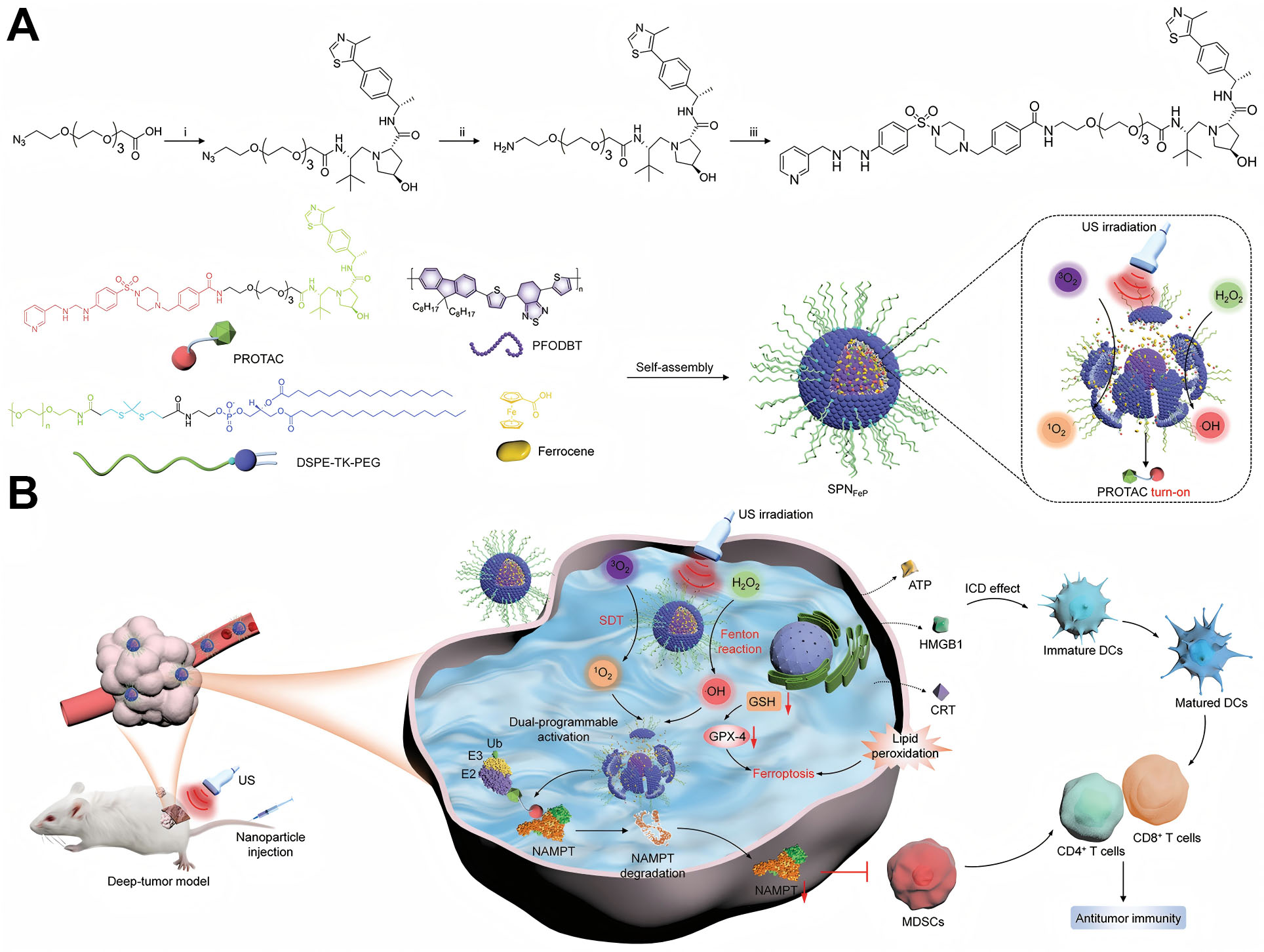
Figure 6. Design of dual-programmable semiconducting polymer nanoPROTACs (SPNFeP) for activatable combination immunotherapy of deep-tissue tumors. (A) Scheme for preparation of SPNFeP via nano-precipitation and dual- programmable activation mechanism; (B) Scheme for dual-programmable activation of SPNFeP, mechanism of SDT and ferroptosis, immune response activation, and suppression of MDSC expansion for immunotherapy of deep-tissue tumors. Reproduced with permission[123]. Copyright 2024, Small. US: Ultrasound; PROTAC: proteolysis targeting chimera; DSPE-TK-PEG: 1,2-Distearoyl-sn-glycero-3-phosphoethanolamine-Thioketal-Polyethylene Glycol; PFODBT: poly[2,7-(9,9-di-octyl-fluorene)-alt-4,7-bis(thiophen-2-yl)benzo-2,1,3-thiadiazole]; GPX-4: glutathione Peroxidase 4; GSH: glutathione (Reduced Glutathione); HMGB1: high-mobility group box protein 1; CRT: calreticulin; MDSC: myeloid-derived suppressor cell; CD4+: CD8+: cluster of differentiation 8 (positive); SDT: sonodynamic therapy; ROS: reactive oxygen species; NAMPT: nicotinamide phosphoribosyltransferase; NAD+: nicotinamide adenine dinucleotide; ICD: immunogenic cell death.
Ultrasound immunotherapy combined with emerging immunotherapy strategies
In addition to immune checkpoint inhibitors and STING activators, ultrasound-mediated immunotherapy can also work synergistically with emerging immunotherapeutic strategies such as adoptive cell therapy (ACT) and tumor vaccines. ACT includes cell therapies such as CAR-T and CAR-NK[124], whose efficacy in solid tumors is often limited by the immunosuppressive tumor microenvironment and physical barriers[125]. Ultrasound-induced ICD can release large amounts of tumor antigens and inflammatory signals, promoting dendritic cell maturation, enhancing the recruitment of effector T cells, and improving the local immune status of the tumor, thereby creating favorable conditions for ACT cell infiltration and functional exertion. At the same time, some ultrasound-responsive nanoplatforms can further enhance the antitumor activity of cell therapy by regulating tumor metabolism, alleviating hypoxia, and lifting immune suppression. To address these bottlenecks, recent research has developed a chemokine-based injectable navigation system (CITE). This system achieves sustained release by forming a drug reservoir through the peritumoral injection of a thermosensitive immunogel co-loaded with CXCL9-conjugated particles and the PD-1 antibody (aPD1). Simultaneously, intravenous injection of the tumor-penetrating peptide iRGD promotes the accumulation of the gel-released drug within the tumor. On the one hand, this creates a sustained CXCL9 chemotactic gradient at the tumor site, guiding CXCR3-expressing CAR-T/TCR-T cells to migrate specifically to the tumor, thereby maximizing the number of migrated T cells within the tumor. On the other hand, aPD-1 blocks immune suppression pathways to enhance the functional activity of infiltrating T cells [Figure 7].
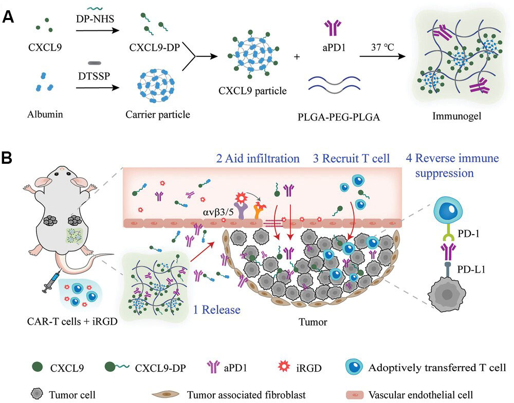
Figure 7. Design of CITE. (A) Flow chart for synthesis of injectable thermo-sensitive immunogel; (B) Schematic diagram illustrating how CITE enhances the therapeutic efficacy of adoptively transferred T cells for solid tumors. (1) Immunogel forms a drug depot and allows sustained release of CXCL9 and PD-1 antibody (aPD1) peritumorally. (2) iRGD, co-infused with transferred T cells, aids tumor infiltration of CXCL9 and aPD1. (3) CXCL9 chemotactic gradient recruits transferred T cells to migrate to and infiltrate the tumor. (4) Intratumoral aPD1 helps tumor-infiltrating T cells to counter immunosuppression. Reproduced with permission[126]. Copyright 2024, Advanced Functional Materials. DP-NHS: Diphosphate-N-Hydroxysuccinimide ester; DTSSP: 3,3'-dithiobis(sulfosuccinimidylpropionate); CXCL9: CXC-chemokine ligand 9; CXCL9-DP: C-X-C motif chemokine ligand 9 - diphenyl (or DP-modified); PLGA: poly(lactic-co-glycolic acid); PEG: poly(ethylene glycol); iRGD: internalizing RGD (peptide); PD-1: programmed cell death protein 1; PD-L1: programmed death-ligand 1; CITE: chemokine-based injectable navigation system; aPD-1: anti-programmed death-1 antibody; CAF: cancer-associated fibroblast.
Furthermore, ultrasound immunotherapy and tumor vaccines exhibit natural synergy. Ultrasound-induced tumor cell death releases abundant tumor-associated antigens (TAAs), neoantigens, and DAMPs such as CRT, ATP, and HMGB1, creating an effect similar to an “in situ vaccine” that significantly enhances antigen presentation efficiency and T-cell activation capacity[127]. In recent years, various ultrasound-responsive nanoplatforms have been used in combination with personalized tumor vaccines or messenger RNA (mRNA) vaccines to achieve a stronger systemic antitumor immune response by simultaneously enhancing antigen release, cross-presentation, and the formation of immune memory.
In summary, although the target molecules and mechanisms of action vary among different combination therapy strategies, they all essentially promote the transition from local immune activation to systemic antitumor immunity by synergistically regulating multiple key components of the Cancer-Immunity Cycle [Figure 8].
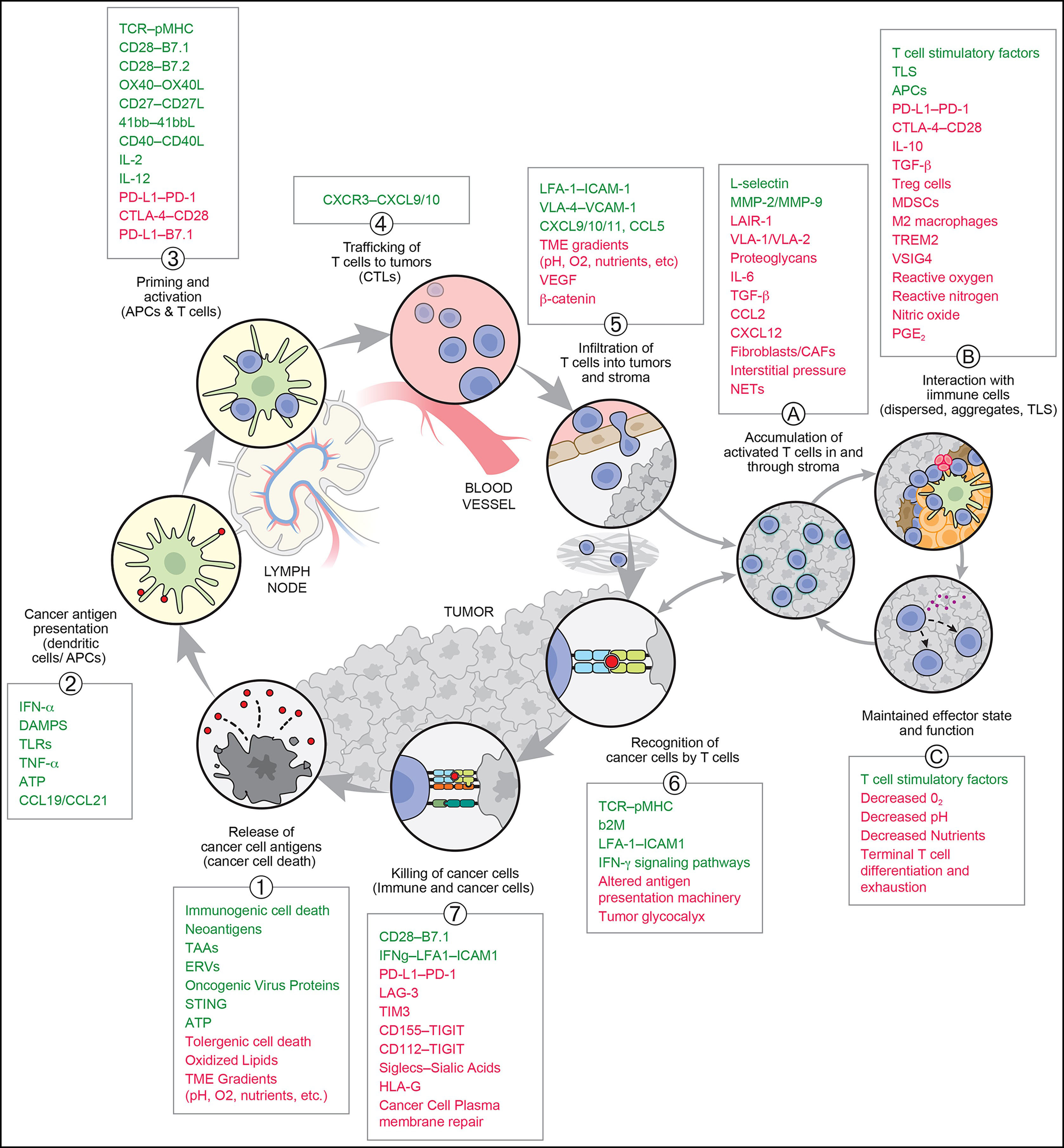
Figure 8. The cancer-immunity cycle with stimulatory and inhibitory factors. Green text denotes stimulatory factors for T cells; red text denotes inhibitory factors. Reproduced with permission[114]. Copyright 2023, Immunity. TCR-pMHC: PD-1: programmed cell death protein 1; PD-L1: programmed death-ligand 1; DAMP: damage-associated molecular pattern; TNF: tumor necrosis factor; CCL: chemokine (C-C motif) ligand; LFA-1: lymphocyte function-associated antigen 1; ICAM1: intercellular cell adhesion molecule 1; IFN: interferon; TME: tumor microenvironment; MDMC: monocyte-derived immune cell; TLS: tertiary lymphoid structure; CTLA: cytotoxic T-lymphocyte-associated protein; TGF: transforming growth factor; TREM2: triggering receptor expressed on myeloid cells 2; VSIG4: V-set and immunoglobulin domain-containing protein 4; ICD: immunogenic cell death; TAA: tumor-associated antigen; ERV: endogenous retrovirus; TLR: toll-like receptor; VEGF: vascular endothelial growth factor; Lair-1: leukocyte-associated immunoglobulin-like receptor-1; b2M: beta-2 microglobulin; LAG-3: lymphocyte-activation gene 3; TIM-3: T-cell immunoglobulin and mucin-domain containing-3; TIGIT: T cell immunoreceptor with Ig and ITIM domains; Siglec-F: sialic acid-binding Ig-like lectin F; APC: antigen-presenting cell; PGE2: prostaglandin E2.
CHALLENGES AND OUTLOOK
In recent years, ultrasound-based immunotherapy enabled by nanomedicine has advanced rapidly in cancer treatment. By harnessing ultrasound-induced ICD, remodeling the tumor immune microenvironment, and systemically activating antitumor immunity, ultrasound-responsive nanoplatforms can effectively promote tumor antigen release, enhance dendritic cell maturation and T-cell infiltration, and exert synergistic effects with immune checkpoint blockade, STING activation, ferroptosis induction, and tumor vaccination. The emergence of diverse nanoplatforms - including organic/inorganic sonosensitizers, piezoelectric nanomaterials, ultrasound-controlled drug delivery vehicles, and biomimetic platforms - has further propelled the field beyond localized tumor ablation toward systemic immune regulation, highlighting its considerable clinical promise.
Despite these advances, several critical challenges remain before clinical translation can be realized. First, the spatiotemporal dynamics of ROS generation and the molecular determinants of ICD induction are not yet fully elucidated, particularly regarding how mechanical indices and duty cycles influence immune phenotype conversion. Second, the long-term biosafety, biodegradability, and in vivo metabolic fate of nanomaterials require systematic evaluation under standardized experimental conditions. Third, the absence of unified ultrasonic parameter protocols introduces variability across preclinical studies, complicating cross-study comparisons and translational benchmarking. Importantly, the biosafety profile of ultrasound itself warrants careful consideration. High-intensity or prolonged ultrasound exposure - especially under elevated mechanical indices - may provoke vascular endothelial injury, increased vascular permeability, and even thrombus formation. Future clinical protocols must therefore define rigorous safety thresholds by incorporating vascular integrity assessments and hemostatic function monitoring into preclinical study designs.
Future research should focus on elucidating the biological mechanisms of ultrasound, establishing a standardized evaluation system and a database of treatment parameters, while simultaneously promoting the development of nanotechnology platforms toward simplicity, scalability, and clinical translational feasibility[128,129]. Leveraging mature clinical equipment platforms such as focused ultrasound and combining them with existing immunotherapy regimens - such as PD-1/PD-L1 antibodies - ultrasound-mediated immunotherapy may hold a favorable position for early clinical exploration as a combination therapy, potentially offering new treatment strategies for the precision treatment and long-term immune control of solid tumors.
With the rapid development of artificial intelligence, precision medicine, and tumor immunology, ultrasound-based immunotherapy is gradually evolving from a localized tumor ablation technique into a systemic immune regulation platform. In the future, artificial intelligence and machine learning are expected to integrate patient tumor characteristics, tissue acoustic parameters, and treatment feedback to enable intelligent optimization of ultrasound parameters and personalized treatment design[130]. At the same time, ultrasound-responsive nanoplatforms will evolve toward multi-stimulus responsiveness, intelligence, and precision. By integrating various tumor microenvironment features - such as pH, ROS, enzymes, and immune signals - these platforms will enable on-demand drug release and dynamic regulation of the treatment process[131].
Beyond single-cell-population targeting, future investigations will increasingly focus on the holistic remodeling of the tumor immune ecosystem. Synergistic regulation of diverse stromal and immune constituents - including tumor-associated macrophages (TAMs), neutrophils (TANs), cancer-associated fibroblasts (CAFs), dendritic cells (DCs), and effector T cells - will be essential to achieve deep and durable immune activation. Notably, inspired by recent advances in two-dimensional materials with exceptional photothermal conversion efficiency, the development of composite nanomaterials integrating near-infrared photothermal responsiveness with sonosensitization represents a compelling frontier. Such “photo-sono” hybrid platforms could leverage photothermal effects to enhance tumor perfusion and alleviate hypoxia - a major limiting factor in ROS-mediated sonodynamic therapies - while simultaneously amplifying ICD through mild hyperthermia[132]. This dual-modality strategy holds promise for breaking the efficacy ceiling of single-mode ultrasound immunotherapy. Ultimately, by integrating cutting-edge technologies such as single-cell sequencing, multi-omics profiling, and digital pathology, it is anticipated that a precision ultrasound immunotherapy framework - tailored to individual patients’ immunological and biomechanical landscapes - will emerge. This trajectory will provide new avenues for personalized cancer care and enduring systemic antitumor immunity[133-135].
CONCLUSIONS
Ultrasound immunotherapy is progressively extending the capabilities of conventional ultrasound technology, shifting from a primarily local tumor ablation modality toward a therapeutic approach with the potential to actively modulate antitumor immune responses. Preclinical studies indicate that ultrasound, particularly when combined with nanomedicine, can induce ICD, promote tumor antigen release, and activate both innate and adaptive immunity, thereby initiating a productive antitumor immune cascade in experimental models. Advances in nanomedicine have further conferred upon this strategy the capacity for targeted drug delivery, tumor microenvironment remodeling, and augmentation of systemic immunity. Over recent years - from the development of sonosensitizers and piezoelectric nanoplatforms to the integration with immune checkpoint blockade, STING activation, ferroptosis induction, and other combinatorial strategies - ultrasound-mediated immunotherapy has demonstrated encouraging potential in preclinical settings to address some of the key limitations of current immunotherapies for solid tumors.
Despite these promising developments, several challenges remain to be addressed before clinical translation. Key issues include the incomplete understanding of underlying mechanistic details, the paucity of long-term biosafety and pharmacokinetic data for nanomaterials, the lack of standardized ultrasound parameter protocols, and the need for robust translational validation. While the deep tissue penetration, excellent spatiotemporal controllability, and inherent compatibility with existing immunotherapies offer a strong rationale for further development, these perceived advantages must be rigorously evaluated in well-designed clinical trials. Looking ahead, continued advances in smart nanomaterials, precision ultrasound modulation, artificial intelligence-assisted treatment optimization, and personalized immunotherapy strategies may enable more refined regulation of the tumor immune ecosystem. Preclinical evidence suggests the possibility of establishing durable systemic antitumor immunity with memory effects, though such outcomes remain to be confirmed in human studies. It is important to emphasize that current expectations regarding clinical utility are largely derived from preclinical findings; extensive translational research will be essential to determine whether ultrasound-mediated immunotherapy can ultimately provide a sound scientific basis and viable clinical paradigm for the precision management of solid tumors.
DECLARATIONS
Authors’ contributions
Made substantial contributions to conception and design of the study and performed data analysis and interpretation: Li J, Liu Y, Hu D
Provided administrative, technical, and material support: Li J
Availability of data and materials
Not applicable.
AI and AI-assisted tools statement
Not applicable.
Financial support and sponsorship
Not applicable.
Conflicts of interest
Li J is an Associate Editor of Nanomedicine Therapeutics. Li J was not involved in any steps of editorial processing, notably including reviewers’ selection, manuscript handling and decision making. The other authors declare that there are no conflicts of interest.
Ethical approval and consent to participate
Not applicable.
Consent for publication
Not applicable.
Copyright
© The Author(s) 2026.
REFERENCES
1. Demidov L, Samoylenko I, Kharkevich G, et al. Improved clinical outcomes with low-dose anti-CTLA-4 (Nurulimab) plus anti-PD-1 (Prolgolimab) vs. anti-PD-1 monotherapy in advanced cutaneous melanoma: results from the phase III OCTAVA trial. Eur J Cancer. 2025;227:115674.
2. Jahangir M, Yazdani O, Kahrizi MS, et al. Clinical potential of PD-1/PD-L1 blockade therapy for renal cell carcinoma (RCC): a rapidly evolving strategy. Cancer Cell Int. 2022;22:401.
3. Wang Q, Li X, Qiu J, et al. A pathway-based mutation signature to predict the clinical outcomes and response to CTLA-4 inhibitors in melanoma. Comput Struct Biotechnol J. 2023;21:2536-46.
4. Lim SY, Shklovskaya E, Lee JH, et al. The molecular and functional landscape of resistance to immune checkpoint blockade in melanoma. Nat Commun. 2023;14:1516.
5. Gambichler T, Finis C, Abu Rached N, et al. Expression of DNA mismatch repair proteins in melanoma patients treated with immune checkpoint inhibitors. J Cancer Res Clin Oncol. 2023;149:1241-7.
6. Hellmann MD, Paz-Ares L, Bernabe Caro R, et al. Nivolumab plus ipilimumab in advanced non-small-cell lung cancer. N Engl J Med. 2019;381:2020-31.
7. Mahoney KM, Ross-Macdonald P, Yuan L, et al. Soluble PD-L1 as an early marker of progressive disease on nivolumab. J Immunother Cancer. 2022;10:e003527.
8. Hu Y, Zhou Y, Zhang M, et al. Genetically modified CD7-targeting allogeneic CAR-T cell therapy with enhanced efficacy for relapsed/refractory CD7-positive hematological malignancies: a phase I clinical study. Cell Res. 2022;32:995-1007.
9. Missiaen R, Lesner NP, Simon MC. HIF: a master regulator of nutrient availability and metabolic cross-talk in the tumor microenvironment. EMBO J. 2023;42:e112067.
10. Jin M, Cao W, Chen B, Xiong M, Cao G. Tumor-derived lactate creates a favorable niche for tumor via supplying energy source for tumor and modulating the tumor microenvironment. Front Cell Dev Biol. 2022;10:808859.
11. Zalpoor H, Aziziyan F, Liaghat M, et al. The roles of metabolic profiles and intracellular signaling pathways of tumor microenvironment cells in angiogenesis of solid tumors. Cell Commun Signal. 2022;20:186.
12. Flies DB, Langermann S, Jensen C, Karsdal MA, Willumsen N. Regulation of tumor immunity and immunotherapy by the tumor collagen extracellular matrix. Front Immunol. 2023;14:1199513.
13. Chen Z, Han F, Du Y, Shi H, Zhou W. Hypoxic microenvironment in cancer: molecular mechanisms and therapeutic interventions. Signal Transduct Target Ther. 2023;8:70.
14. Wang T, Wang J, Jiang H, et al. Targeted regulation of tumor microenvironment through the inhibition of MDSCs by curcumin loaded self-assembled nano-filaments. Mater Today Bio. 2022;15:100304.
15. Sattiraju A, Kang S, Giotti B, et al. Hypoxic niches attract and sequester tumor-associated macrophages and cytotoxic T cells and reprogram them for immunosuppression. Immunity. 2023;56:1825-1843.e6.
16. Hong M, Talluri S, Chen YY. Advances in promoting chimeric antigen receptor T cell trafficking and infiltration of solid tumors. Curr Opin Biotechnol. 2023;84:103020.
17. Ma L, Hostetler A, Morgan DM, et al. Vaccine-boosted CAR T crosstalk with host immunity to reject tumors with antigen heterogeneity. Cell. 2023;186:3148-3165.e20.
18. Wang Y, Drum DL, Sun R, et al. Stressed target cancer cells drive nongenetic reprogramming of CAR T cells and solid tumor microenvironment. Nat Commun. 2023;14:5727.
19. Cao Z, Li D, Zhao L, et al. Bioorthogonal in situ assembly of nanomedicines as drug depots for extracellular drug delivery. Nat Commun. 2022;13:2038.
20. Chen Y, Huang Y, Li Q, et al. Targeting Xkr8 via nanoparticle-mediated in situ co-delivery of siRNA and chemotherapy drugs for cancer immunochemotherapy. Nat Nanotechnol. 2023;18:193-204.
21. Li J, Luo Y, Zeng Z, et al. Precision cancer sono-immunotherapy using deep-tissue activatable semiconducting polymer immunomodulatory nanoparticles. Nat Commun. 2022;13:4032.
22. Liu L, Pan D, Chen S, et al. Systematic design of cell membrane coating to improve tumor targeting of nanoparticles. Nat Commun. 2022;13:6181.
23. Zelepukin IV, Griaznova OY, Shevchenko KG, et al. Flash drug release from nanoparticles accumulated in the targeted blood vessels facilitates the tumour treatment. Nat Commun. 2022;13:6910.
24. Hao Y, Gao Y, Fan Y, et al. A tumor microenvironment-responsive poly(amidoamine) dendrimer nanoplatform for hypoxia-responsive chemo/chemodynamic therapy. J Nanobiotechnology. 2022;20:43.
25. Song J, Cheng M, Xie Y, Li K, Zang X. Efficient tumor synergistic chemoimmunotherapy by self-augmented ROS-responsive immunomodulatory polymeric nanodrug. J Nanobiotechnology. 2023;21:93.
26. Tang J, Zhang X, Cheng L, et al. Multiple stimuli-responsive nanosystem for potent, ROS-amplifying, chemo-sonodynamic antitumor therapy. Bioact Mater. 2022;15:355-71.
27. Tang M, Chen B, Xia H, et al. pH-gated nanoparticles selectively regulate lysosomal function of tumour-associated macrophages for cancer immunotherapy. Nat Commun. 2023;14:5888.
28. Wang Y, Wang Q, Wang X, et al. Docetaxel-loaded pH/ROS dual-responsive nanoparticles with self-supplied ROS for inhibiting metastasis and enhancing immunotherapy of breast cancer. J Nanobiotechnology. 2023;21:286.
29. Mao D, Li B, Li J, Pan J, Wu Z, Feng N. Nanodrug delivery systems in tumor immunotherapy: From immune activation to tumor reprogramming. Adv Drug Deliv Rev. 2026;232:115838.
30. He J, Xue W, Li Y. Tumor microenvironment-responsive nanomedicine: monitoring and modulating the tumor microenvironment for precision cancer therapy. Int J Nanomedicine. 2026;21:560983.
31. Jia Q, Wang A, Yuan Y, Zhu B, Long H. Heterogeneity of the tumor immune microenvironment and its clinical relevance. Exp Hematol Oncol. 2022;11:24.
32. Cressey P, Abd Shukor SB, Thanou M. Sonodynamic therapy: transforming sound into light for hard-to-treat tumours. Adv Drug Deliv Rev. 2025;226:115696.
33. Yuan F, Peng D, Lu M, Zhang K, Mi P, Xu J. Ultrasound-responsive nanocarriers for cancer therapy: Physiochemical features-directed design. J Control Release. 2025;388:114353.
34. Bano I, Jorratt P, Kútna V, Pala J, Tsenov G. Noninvasive focused ultrasound as a safe modulator of calcium-dependent neurochemical signalling in primary cortical cultures. Neurochem Res. 2026;51:61.
35. Kojima Y. Effects of low-intensity pulsed ultrasound on muscle mass and Fndc5 mRNA expression in aged male mice. Biogerontology. 2025;26:187.
36. Ayyami Y, Dastgir M, Yektamanesh M, et al. Advances in ultrasound-activated nano-sonosensitizers for cancer treatment: a systematic review and meta-analysis. Ultrason Sonochem. 2026;127:107750.
37. Kong C, Luo C, Xue F, et al. Ultrasound-triggered charge-reversal nanoparticles via golgi-dependent iterative transcytosis for enhanced deep tumor penetration. ACS Nano. 2026;20:4741-57.
38. Liang G, Sadhukhan T, Banerjee S, et al. Reduction of platinum(IV) prodrug hemoglobin nanoparticles with deeply penetrating ultrasound radiation for tumor-targeted therapeutically enhanced anticancer therapy. Angew Chem Int Ed Engl. 2023;62:e202301074.
39. Chen Y, Du M, Yuan Z, Chen Z, Yan F. Spatiotemporal control of engineered bacteria to express interferon-γ by focused ultrasound for tumor immunotherapy. Nat Commun. 2022;13:4468.
40. Fabiano AR, Newman MW, Dombroski JA, et al. Applying ultrasound to mechanically and noninvasively sensitize prostate tumors to TRAIL-mediated apoptosis. Adv Sci (Weinh). 2025;12:e2412995.
41. Pepple AL, Guy JL, McGinnis R, et al. Spatiotemporal local and abscopal cell death and immune responses to histotripsy focused ultrasound tumor ablation. Front Immunol. 2023;14:1012799.
42. Wu Y, Huang Z, Liu Y, et al. Ultrasound control of genomic regulatory toolboxes for cancer immunotherapy. Nat Commun. 2024;15:10444.
43. He J, Liu Z, Zhu X, Xia H, Gao H, Lu J. Ultrasonic microbubble cavitation enhanced tissue permeability and drug diffusion in solid tumor therapy. Pharmaceutics. 2022;14:1642.
44. Imran KM, Tintera B, Morrison HA, et al. Improved therapeutic delivery targeting clinically relevant orthotopic human pancreatic tumors engrafted in immunocompromised pigs using ultrasound-induced cavitation: a pilot study. Pharmaceutics. 2023;15:1585.
45. Luo Y, Qiao B, Yang C, et al. Low intensity focused ultrasound ignited “deep-penetration nanobomb” (DPNB) for tetramodal imaging guided hypoxia-tolerant sonodynamic therapy against hypoxic tumors. Int J Nanomedicine. 2022;17:4547-65.
46. Ye D, Wang G, Liu X, et al. Cascade ultrasonic cavitation enables microbubble-nanoparticle hybrid to broadly accumulate and penetrate into pancreatic ductal adenocarcinoma. Small Structures. 2025;6:2500077.
47. Chen Y, Song Y, Zhang C, et al. Ultrasound-responsive release of CD39 inhibitor overcomes adenosine-mediated immunosuppression in triple-negative breast cancer. J Control Release. 2025;383:113819.
48. Fang M, Zheng J, Song Q, et al. Breaking apoptosis-induced immune silence: ultrasound-activated nano-oncolytic therapy reinvigorates antitumor immunity. Adv Mater. 2025;37:e2508681.
49. Yang J, Liao M, Wu Z, et al. Perfluorohexane nanodroplet-assisted mechanical high intensity focused ultrasound cavitation: a strategy for hepatocellular carcinoma treatment. Acta Biomater. 2025;195:297-308.
50. Bhalotia A, Hutchinson DW, Kosmides T, et al. Enhanced delivery of lipid nanoparticle-based immunotherapy by modulating the tumor tissue stiffness using ultrasound-activated nanobubbles. ACS Nano. 2026;20:4592-606.
51. Chen L, Liu J, Chen Q, et al. Modulating tumor interstitial fluid pressure using ultrasound and microbubble therapy: a preclinical study for enhanced drug delivery in cancer treatment. BMC Cancer. 2025;26:40.
52. Yang G, Li H, Yin J, et al. Alleviating tumor hypoxia and immunosuppression via sononeoperfusion: a new ally for potentiating anti-PD-L1 blockade of solid tumor. Ultrason Sonochem. 2025;112:107115.
53. Jiang Z, Xiao W, Fu Q. Stimuli responsive nanosonosensitizers for sonodynamic therapy. J Control Release. 2023;361:547-67.
54. Chen F, Zhang H, Li S, et al. Polypeptide sono-adjuvant for ultrasound-activatable regulation of innate immunity and cancer vaccination therapy. Nat Commun. 2025;17:272.
55. Tang J, Chen Y, Lai H, et al. Innovative immunosonogen nanoplatform combining sonodynamic oxygenation and immunomodulation for superior cancer therapy. Chem Eng J. 2025;515:163582.
56. Xue H, Jin J, Huang X, et al. Wearable flexible ultrasound microneedle patch for cancer immunotherapy. Nat Commun. 2025;16:2650.
57. Yang M, Zhang W, Su L, et al. Biomimetic nanoadjuvant-enhanced ultrasound-induced STING activation potentiates aPD-L1 therapy to overcome cancer recurrence and metastasis. Chem Eng J. 2025;511:161735.
58. Ma K, Dong J, Chen Z, et al. Ultrasound-powered piezocatalytic nanomotors with dual-mode synergistic/cascade propulsion reprogram hypoxic tumor-associated macrophages for enhanced cancer immunotherapy. Chem Eng J. 2025;512:162107.
59. Wang C, Zhang R, He J, et al. Ultrasound-responsive low-dose doxorubicin liposomes trigger mitochondrial DNA release and activate cGAS-STING-mediated antitumour immunity. Nat Commun. 2023;14:3877.
60. Ren S, Zhang M, Cai C, et al. A carrier-free ultrasound-responsive polyphenol nanonetworks with enhanced sonodynamic-immunotherapy for synergistic therapy of breast cancer. Biomaterials. 2025;317:123109.
61. McCorkell G, Nakayama M, Feltis B, Piva T, Geso M. Ultrasound-stimulated microbubbles enhance radiation-induced cell killing. Ultrasound Med Biol. 2022;48:2449-60.
62. Du JR, Wang Y, Yue ZH, et al. Recent advances in sonodynamic immunotherapy. J Cancer Res Clin Oncol. 2023;149:1645-56.
63. Tao Y, Zhuang W, Fan W, et al. Dual-functional silver nanoparticle-enhanced ZnO nanorods for improved reactive oxygen species generation and cancer treatment. iScience. 2025;28:111858.
64. Zhaoyun L, Wang H, Yang C, et al. Enhancing antitumor immunity via ROS-ERS and pyroptosis-induced immunogenic cell death in multiple myeloma. J Immunother Cancer. 2025;13:e011717.
65. Fite BZ, Wang J, Kare AJ, et al. Immune modulation resulting from MR-guided high intensity focused ultrasound in a model of murine breast cancer. Sci Rep. 2021;11:927.
66. Akter S, Madhuvilakku R, Kar AK, et al. Reactive oxygen species (ROS) in cancer: from mechanism to therapeutic implications. Signal Transduct Target Ther. 2026;11.
67. Wu X, Liang J, Shu J, et al. Narrow-bandgap iridium(III)-C3N5 nanocomplex as an oxygen self-sufficient piezo-sonosensitizer for hypoxic tumor sonodynamic immunotherapy. J Am Chem Soc. 2025;147:15329-43.
68. Zhang L, Yi H, Song J, et al. Mitochondria-targeted and ultrasound-activated nanodroplets for enhanced deep-penetration sonodynamic cancer therapy. ACS Appl Mater Interfaces. 2019;11:9355-66.
69. Lei Q, Zhou W, Gao S, et al. Ultrasound-responsive metal-organic framework-based nanosystem for sonodynamic therapy/amplified ferroptosis/IDO-blockade osteosarcoma immunotherapy. Chem Eng J. 2024;490:151614.
70. Wang Q, He J, Qi Y, Ye Y, Ye J, Zhou M. Ultrasound-enhanced nano catalyst with ferroptosis-apoptosis combined anticancer strategy for metastatic uveal melanoma. Biomaterials. 2024;305:122458.
71. Zheng H, Lin H, Ke Y, et al. Piezoelectric-triggered ferroptosis for cancer therapy via cascade-enhanced immune response. Adv Healthc Mater. 2025;14:e2501339.
72. Rucker AJ, Park CS, Li QJ, Moseman EA, Chan FK. Necroptosis stimulates interferon-mediated protective anti-tumor immunity. Cell Death Dis. 2024;15:403.
73. Mannion J, Gifford V, Bellenie B, et al. A RIPK1-specific PROTAC degrader achieves potent antitumor activity by enhancing immunogenic cell death. Immunity. 2024;57:1514-1532.e15.
74. Zuo Z, Yin H, Zhang Y, Xie C, Wang Q. A cytotoxic T cell inspired oncolytic nanosystem promotes lytic cell death by lipid peroxidation and elicits antitumor immune responses. Nat Commun. 2023;14:5456.
75. Lv Z, Liu Z, Nie Z, et al. Ferroptosis reshapes the tumor immune microenvironment: molecular mechanisms, immune regulation, and therapeutic synergistic strategies. Front Immunol. 2026;17:1772259.
76. Li M, Liu Y, Zhang Y, Yu N, Li J. Sono-activatable semiconducting polymer nanoreshapers multiply remodel tumor microenvironment for potent immunotherapy of orthotopic pancreatic cancer. Adv Sci (Weinh). 2023;10:e2305150.
77. Gao J, Wang WQ, Pei Q, Lord MS, Yu HJ. Engineering nanomedicines through boosting immunogenic cell death for improved cancer immunotherapy. Acta Pharmacol Sin. 2020;41:986-94.
78. Humeau J, Sauvat A, Cerrato G, et al. Inhibition of transcription by dactinomycin reveals a new characteristic of immunogenic cell stress. EMBO Mol Med. 2020;12:e11622.
79. Liu P, Zhao L, Pol J, et al. Crizotinib-induced immunogenic cell death in non-small cell lung cancer. Nat Commun. 2019;10:1486.
80. Krysko DV, Garg AD, Kaczmarek A, Krysko O, Agostinis P, Vandenabeele P. Immunogenic cell death and DAMPs in cancer therapy. Nat Rev Cancer. 2012;12:860-75.
81. McNulty S, Colaco CA, Blandford LE, Bailey CR, Baschieri S, Todryk S. Heat-shock proteins as dendritic cell-targeting vaccines - getting warmer. Immunology. 2013;139:407-15.
82. Shen X, Fan S, He J, et al. Designer solid self-emulsifying nanovaccines enable dual modulation of dendritic cells and t cells for potent antitumor immunity. Adv Sci (Weinh). 2026;13:e12139.
83. Chen G, Wang Y, Mo L, et al. Ultrasound-activatable in situ vaccine for enhanced antigen self- and cross-presentation to overcome cancer immunotherapy resistance. ACS Nano. 2024:20296-312.
84. Zhou Y, Shen G, Zhou X, Li J. Therapeutic potential of tumor-associated neutrophils: dual role and phenotypic plasticity. Signal Transduct Target Ther. 2025;10:178.
85. Wang Y, Ma J, Liu Y, et al. Unraveling the complex role of tumor-associated neutrophils within solid tumors. Cancer Immunol Immunother. 2025;74:210.
86. Gao X, Xu M, Xiao H, et al. Tumor-associated neutrophils: a complex role in cancer. Clin Immunol. 2025;280:110558.
87. Huang X, Nepovimova E, Adam V, et al. Neutrophils in cancer immunotherapy: friends or foes? Mol Cancer 2024;23:107.
88. Michelet X, Dyck L, Hogan A, et al. Metabolic reprogramming of natural killer cells in obesity limits antitumor responses. Nat Immunol. 2018;19:1330-40.
89. Myers JA, Miller JS. Exploring the NK cell platform for cancer immunotherapy. Nat Rev Clin Oncol. 2021;18:85-100.
90. Kranz LM, Diken M, Haas H, et al. Systemic RNA delivery to dendritic cells exploits antiviral defence for cancer immunotherapy. Nature. 2016;534:396-401.
91. Raskov H, Orhan A, Christensen JP, Gögenur I. Cytotoxic CD8+ T cells in cancer and cancer immunotherapy. Br J Cancer. 2021;124:359-67.
92. Borst J, Ahrends T, Bąbała N, Melief CJM, Kastenmüller W. CD4+ T cell help in cancer immunology and immunotherapy. Nat Rev Immunol. 2018;18:635-47.
93. Oh DY, Fong L. Cytotoxic CD4+ T cells in cancer: expanding the immune effector toolbox. Immunity. 2021;54:2701-11.
94. Topchyan P, Lin S, Cui W. The role of CD4 T cell help in CD8 T cell differentiation and function during chronic infection and cancer. Immune Netw. 2023;23:e41.
95. Lee JM. Cancer sono-immunotherapy using a multi-metal-ligand framework. Commun Mater. 2024;5:671.
96. Oliveira G, Stromhaug K, Klaeger S, et al. Phenotype, specificity and avidity of antitumour CD8+ T cells in melanoma. Nature. 2021;596:119-25.
97. Rosato PC, Wijeyesinghe S, Stolley JM, Masopust D. Integrating resident memory into T cell differentiation models. Curr Opin Immunol. 2020;63:35-42.
98. Yu N, Zhou J, Ding M, Li M, Peng S, Li J. Sono-triggered cascade lactate depletion by semiconducting polymer nanoreactors for cuproptosis-immunotherapy of pancreatic cancer. Angew Chem Int Ed Engl. 2024;63:e202405639.
99. Guo Y, Wang SZ, Zhang X, et al. In situ generation of micrometer-sized tumor cell-derived vesicles as autologous cancer vaccines for boosting systemic immune responses. Nat Commun. 2022;13:6534.
100. Hirschhorn D, Budhu S, Kraehenbuehl L, et al. T cell immunotherapies engage neutrophils to eliminate tumor antigen escape variants. Cell. 2023;186:1432-1447.e17.
101. Zhang C, Huang J, Zeng Z, et al. Catalytical nano-immunocomplexes for remote-controlled sono-metabolic checkpoint trimodal cancer therapy. Nat Commun. 2022;13:3468.
102. Wang HH, Nan LY, Zheng Y, Guo J. Porphyrin oriented bimetallic sonodynamic MOF for enhanced ferroptosis and cGAS-STING pathway mediated antitumor immunotherapy. J Nanobiotechnology. 2026;24.
103. Zeng J, Yan Z, Wang D, et al. Mitochondria-targeted MXene@MnO2-TPP nanoheterostructures for synergistic enhancement of sonodynamic therapy and immunotherapy in osteosarcoma. Bioact Mater. 2025;54:450-65.
104. Li J, Yue Z, Tang M, et al. Strategies to reverse hypoxic tumor microenvironment for enhanced sonodynamic therapy. Adv Healthc Mater. 2024;13:e2302028.
105. Xu M, Hu Y, Wu J, Liu J, Pu K. Sonodynamic nano-LYTACs reverse tumor immunosuppressive microenvironment for cancer immunotherapy. J Am Chem Soc. 2024;146:34669-80.
106. Feng Z, Wang Z, Xiang X, et al. Progress in nanomedicine for sonodynamic immunotherapy of tumors. EngMedicine. 2024;1:100027.
107. Knight Z, Ruiz A, Elies J. Piezoelectric nanomaterials for cancer therapy: current research and future perspectives on glioblastoma. J Funct Biomater. 2025;16:114.
108. Ya Z, Guo S, Li Y, et al. Focused acoustic vortex-mediated sonochemotherapy for the amplification of immunogenic cell death combined with checkpoint blockade to potentiate cancer immunotherapy. Biomaterials. 2023;301:122278.
109. Li Y, Chen W, Kang Y, et al. Nanosensitizer-mediated augmentation of sonodynamic therapy efficacy and antitumor immunity. Nat Commun. 2023;14:6973.
110. Choi Y, Jung K. Normalization of the tumor microenvironment by harnessing vascular and immune modulation to achieve enhanced cancer therapy. Exp Mol Med. 2023;55:2308-19.
111. Mai Z, Lin Y, Lin P, Zhao X, Cui L. Modulating extracellular matrix stiffness: a strategic approach to boost cancer immunotherapy. Cell Death Dis. 2024;15:307.
112. Sahai E, Astsaturov I, Cukierman E, et al. A framework for advancing our understanding of cancer-associated fibroblasts. Nat Rev Cancer. 2020;20:174-86.
113. Yuan J, Ye D, Chen S, Chen H. Therapeutic ultrasound-enhanced immune checkpoint inhibitor therapy. Front Phys. 2021;9:636985.
114. Mellman I, Chen DS, Powles T, Turley SJ. The cancer-immunity cycle: indication, genotype, and immunotype. Immunity. 2023;56:2188-205.
115. Yu N, Li M, Zhang Y, et al. Dual-modulation of immunosuppressive pathways using sono-activatable semiconducting polymer nanofeedbacks for cancer immunotherapy. Nano Today. 2023;52:101944.
116. Sharma P, Goswami S, Raychaudhuri D, et al. Immune checkpoint therapy-current perspectives and future directions. Cell. 2023;186:1652-69.
117. Tselikas L, Susini S, Texier M, et al. Safety and efficacy of intratumoural anti-CTLA4 with intravenous anti-PD1. Nature. 2026;655:219-29.
118. Corrales L, McWhirter SM, Dubensky TW Jr, Gajewski TF. The host STING pathway at the interface of cancer and immunity. J Clin Invest. 2016;126:2404-11.
119. Lei J, Zhang W, Ma L, et al. Sonodynamic amplification of cGAS-STING activation by cobalt-based nanoagonist against bone and metastatic tumor. Biomaterials. 2023;302:122295.
120. Wang W, Hu Q, Wu C, et al. A ferroptosis-based intelligent nanoplatform with chemo-sonodynamic therapy carrying oxygen for improving tumor suppression and antitumor immunity. J Nanobiotechnology. 2026;24.
122. Yin Y, Jiang X, Sun L, et al. Continuous inertial cavitation evokes massive ROS for reinforcing sonodynamic therapy and immunogenic cell death against breast carcinoma. Nano Today. 2021;36:101009.
123. Wang F, Dong G, Ding M, Yu N, Sheng C, Li J. Dual-programmable semiconducting polymer NanoPROTACs for deep-tissue sonodynamic-ferroptosis activatable immunotherapy. Small. 2024;20:e2306378.
124. Hou AJ, Chen LC, Chen YY. Navigating CAR-T cells through the solid-tumour microenvironment. Nat Rev Drug Discov. 2021;20:531-50.
125. Anderson KG, Stromnes IM, Greenberg PD. Obstacles posed by the tumor microenvironment to T cell activity: a case for synergistic therapies. Cancer Cell. 2017;31:311-25.
126. Wan W, Han W, Chen J, et al. Chemokine-based injectable navigation system for enhancing CAR‐T cell therapy against solid tumors. Adv Funct Mater 2024;34:2401161.
127. Hu Z, Ott PA, Wu CJ. Towards personalized, tumour-specific, therapeutic vaccines for cancer. Nat Rev Immunol. 2018;18:168-82.
128. Labib S, Bright RK, Liu J. Focused ultrasound in cancer immunotherapy: a review of mechanisms and applications. Ultrasound Med Biol. 2025;51:1-14.
129. Zhang P, Xiao Y, Sun X, et al. Cancer nanomedicine toward clinical translation: obstacles, opportunities, and future prospects. Med. 2023;4:147-67.
130. Sadeghi-Goughari M, Rajabzadeh H, Han JW, Kwon HJ. Artificial intelligence-assisted ultrasound-guided focused ultrasound therapy: a feasibility study. Int J Hyperthermia. 2023;40:2260127.
131. Zhang Y, Li J, Pu K. Recent advances in dual- and multi-responsive nanomedicines for precision cancer therapy. Biomaterials. 2022;291:121906.
132. Xie Z, Duo Y, Fan T, et al. Light-induced tumor theranostics based on chemical-exfoliated borophene. Light Sci Appl. 2022;11:324.
133. Baxi V, Edwards R, Montalto M, Saha S. Digital pathology and artificial intelligence in translational medicine and clinical practice. Mod Pathol. 2022;35:23-32.
134. Binnewies M, Roberts EW, Kersten K, et al. Understanding the tumor immune microenvironment (TIME) for effective therapy. Nat Med. 2018;24:541-50.
Cite This Article
 Open Access
How to Cite
Download Citation
Export Citation File:
Type of Import
Tips on Downloading Citation
Citation Manager File Format
Type of Import
Direct Import: When the Direct Import option is selected (the default state), a dialogue box will give you the option to Save or Open the downloaded citation data. Choosing Open will either launch your citation manager or give you a choice of applications with which to use the metadata. The Save option saves the file locally for later use.
Indirect Import: When the Indirect Import option is selected, the metadata is displayed and may be copied and pasted as needed.
About This Article
Copyright

Data & Comments
Data
0





Comments
Comments must be written in English. Spam, offensive content, impersonation, and private information will not be permitted. If any comment is reported and identified as inappropriate content by OAE staff, the comment will be removed without notice. If you have any queries or need any help, please contact us at [email protected].