


fig3
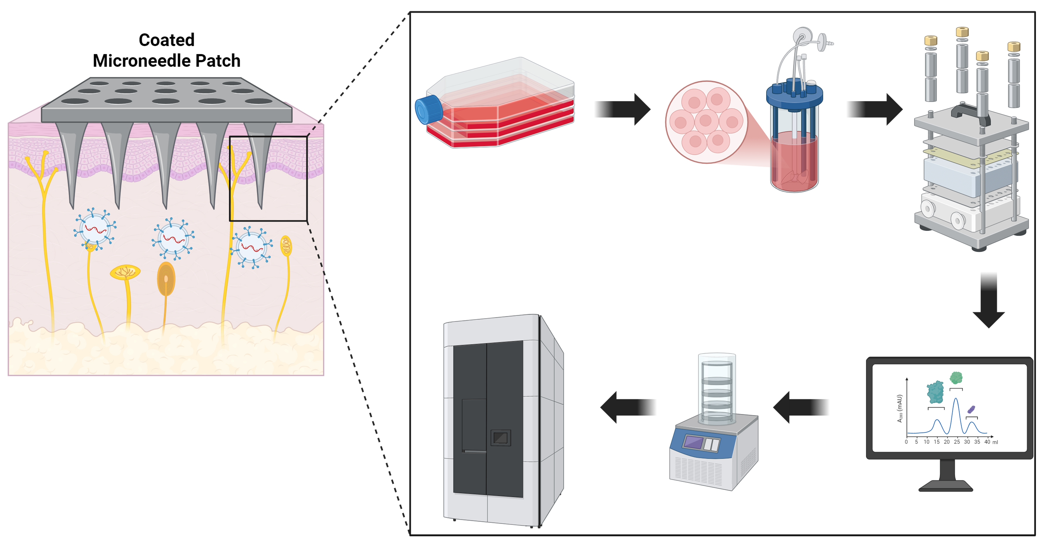
Figure 3. Transdermal delivery and scalable manufacturing/quality control of ADSC-derived exosomes. Left: A coated microneedle patch breaches the stratum corneum and deposits exosomes into the viable epidermis/papillary dermis for uptake by keratinocytes and fibroblasts. Right (workflow): (1) Culture/priming of ADSCs; (2) expansion in a microcarrier or hollow-fiber bioreactor; (3) TFF for clarification and volume reduction (e.g., 300 kDa cassette); (4) size-exclusion chromatography (SEC/qEV or FPLC) for polishing; (5) lyophilization of the exosome-coated microneedle formulation with protectants (e.g., trehalose/sucrose ± surfactant); (6) labeled storage (2-8 °C liquid or -80 °C frozen). Quality control is applied across steps, including NTA (size/count), TEM, EV markers (CD63/CD81/CD9, TSG101), and assessment of protein/lipoprotein carryover. Created with BioRender.com. ADSC: Adipose-derived stem cell; TFF: tangential flow filtration; SEC: size-exclusion chromatography; FPLC: fast protein liquid chromatography; NTA: nanoparticle tracking analysis; TEM: transmission electron microscopy; EV: extracellular vesicle; CD: cluster of differentiation; TSG101: tumor susceptibility gene 101.


