


fig3
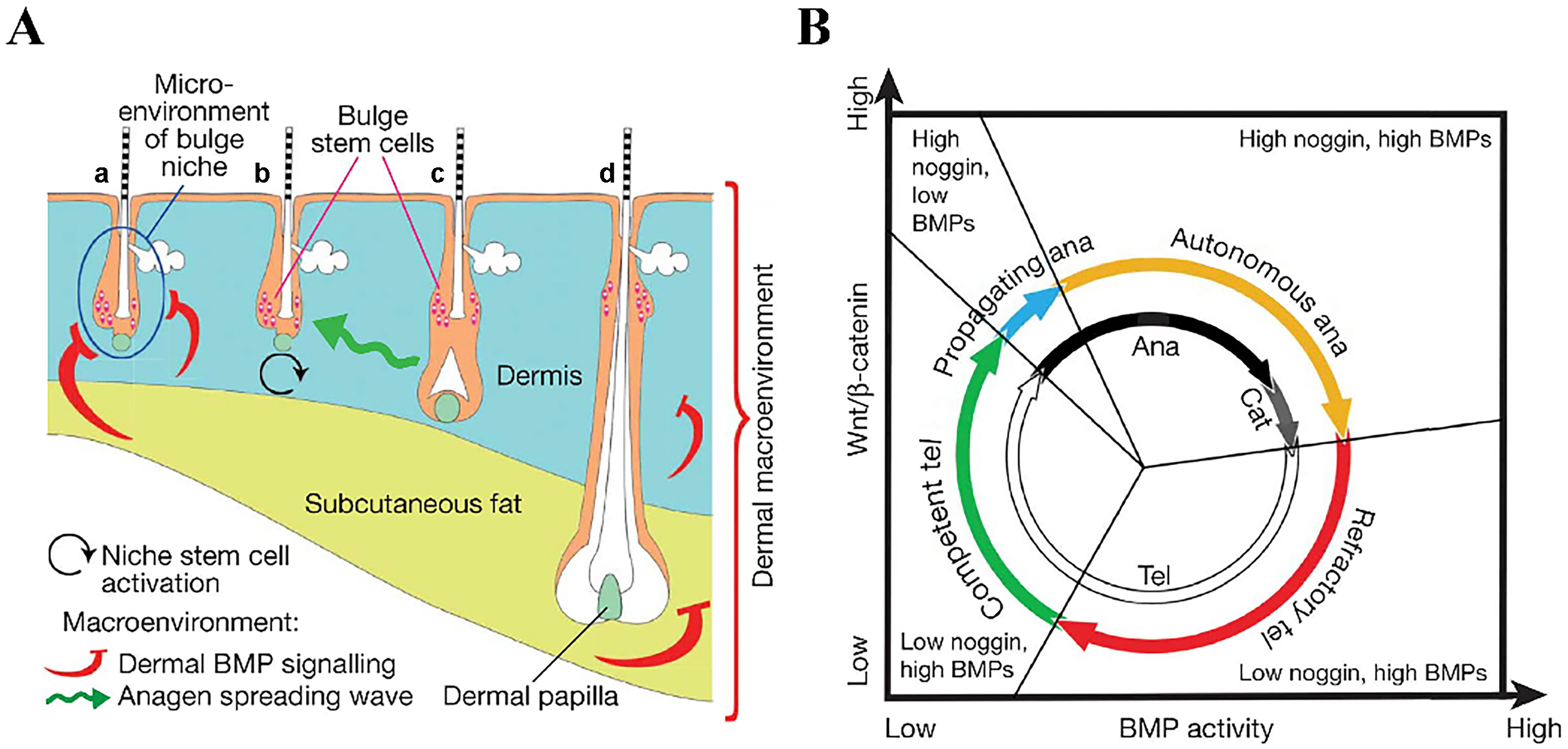
Figure 3. Schematic diagram of the hair growth cycle phases[86]. (A) Illustration of the bulge niche microenvironment and the interfollicular dermal macroenvironment, including the dermis, subcutaneous fat, and adjacent follicles. Arrows indicate anagen-promoting (black and green) or anagen-inhibiting (red) activities. Hair follicles are shown in different phases: (a) refractory telogen; (b) competent telogen; (c) propagating anagen; and (d) autonomous anagen. The blue circle in (A) marks the intrafollicular microenvironment, color-coded to match (B); (B) New functional phases (outer colored circle) mapped onto classical hair-cycle stages (inner black-and-white circle). Based on their ability to induce growth, anagen is subdivided into propagating (blue, inducing) and autonomous (yellow, non-inducing) phases. Telogen is divided into refractory (red) and competent (green) phases according to follicular responsiveness to regenerative signals. BMPs: Bone morphogenetic proteins.


