


fig5
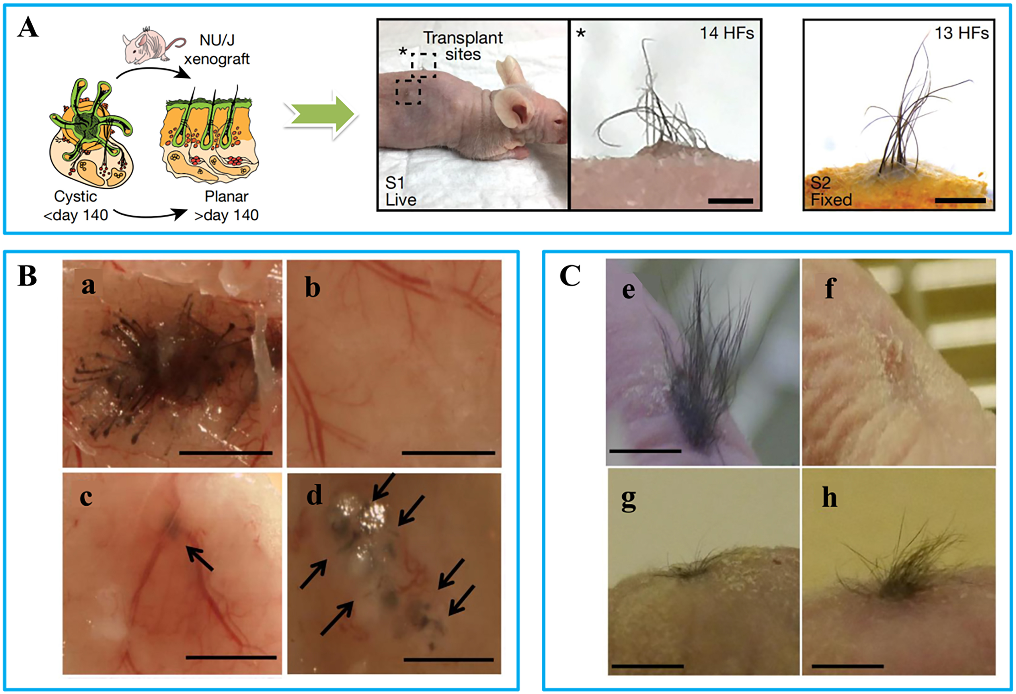
Figure 5. Representative images of different hair transplantation approaches: (A) Skin organ transplantation: images of in vitro cultured skin organoids and hair regeneration before and after transplantation into nude mice[111]; (B) Injection method[112]: (a) Positive controls: freshly isolated dermal and epidermal cells from mouse embryos (E17.5-18.5) were injected; (b) Negative control: only epidermal cells were injected; (c) DPCs treated with FGF2 did not always form hair shafts; (d) DPCs treated with FGF2 + PDGF-AA (10-50 ng/mL) formed hair shafts; (C) Chamber method[112]: (e) Positive control induced hair follicles; (f) Negative control did not induce any follicles; (g) DPCs treated with FGF2 induced sparse hair follicles; (h) DPCs treated with FGF2 + PDGF-AA (20 ng/mL) induced more abundant hair follicles (h). (Scale bar = 200 μm). DPCs: Dermal papilla cells; FGF2: fibroblast growth factor-2; PDGF: platelet-derived growth factor; AA: alopecia areata.


