


fig4
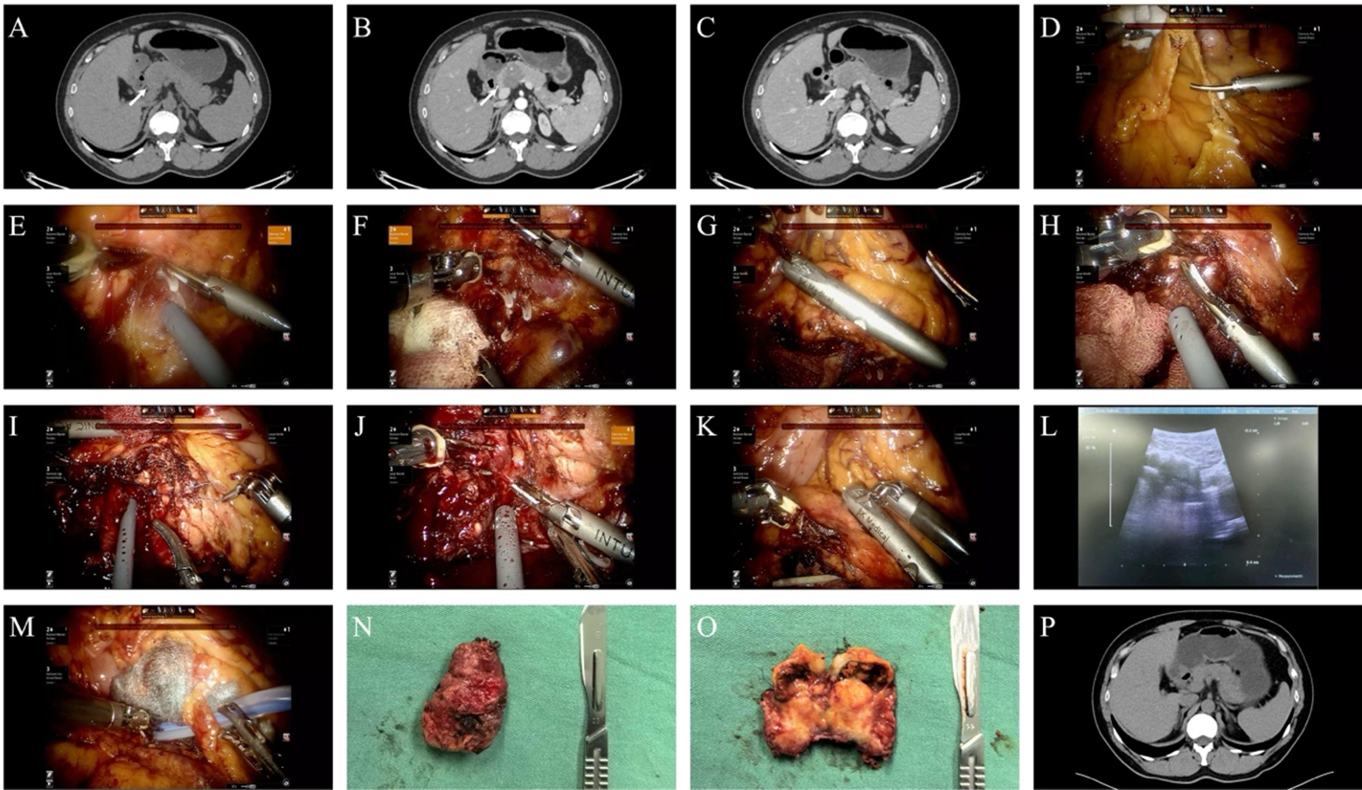
Figure 4. Representative robotic enucleation for a solid pseudopapillary neoplasm of the pancreas. (A-C) Contrast-enhanced computed tomography demonstrating a heterogeneous, relatively hypodense lesion at the pancreatic head–neck junction, measuring approximately 3.8 × 3.0 cm in maximal diameter, with focal calcifications, relative hypoenhancement, and no dilation of the main pancreatic duct; (D) Adhesiolysis; (E) Clip ligation and division of encountered ductal structures; (F) Opening of the gastrocolic ligament to expose the pancreas; (G) Intraoperative ultrasonography revealing a heterogeneous hypoechoic lesion; (H) Stepwise transection of pancreatic parenchyma using an ultrasonic scalpel; (I) Suture ligation of arterial bleeding; (J) Complete enucleation of the lesion with preservation of surrounding pancreatic parenchyma; (K and L) Repeat intraoperative ultrasonography confirming integrity of the main pancreatic duct; (M) Placement of hemostatic materials and surgical drainage; (N and O) Gross appearance of the resected specimen; (P) Postoperative computed tomography showing no abnormal findings and no pancreatic duct dilation.