





fig1
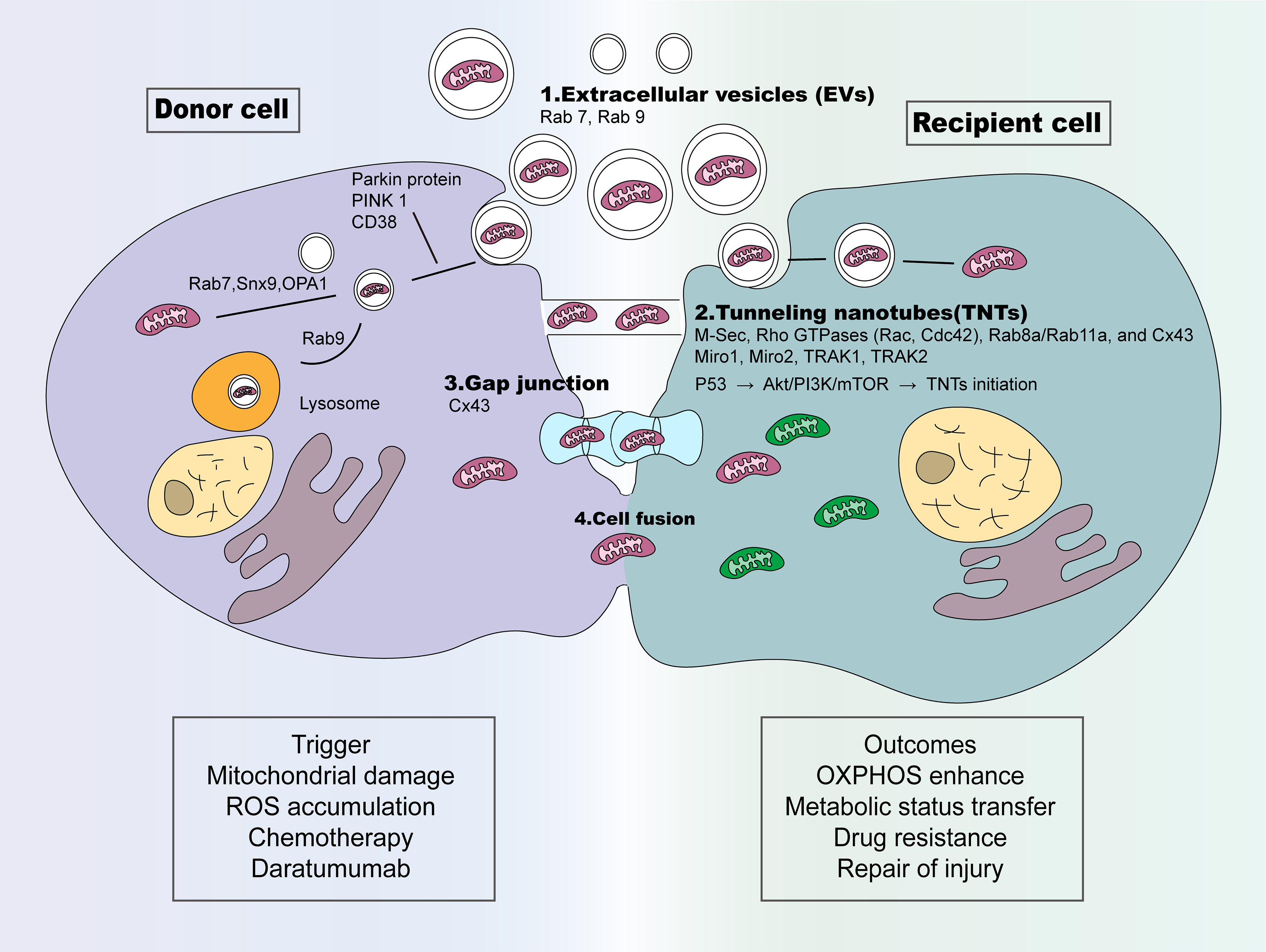
Figure 1. The ways of mitochondrial transfer in cancer cells. Four primary mechanisms underlying mitochondrial transfer have been identified: (1) EVs: Mitochondria-containing EVs form under the regulation of Snx9, Rab9 and OPA1, while Rab7 mediates EVs-lysosome fusion for cargo degradation. Alternatively, PINK1/Parkin signaling drives extracellular release of mitochondrial cargo-laden vesicles; (2) TNTs: Multiple signaling molecules coordinate TNT formation, including M-Sec, Rho GTPases (Rac, Cdc42), Rab8a/Rab11a, and Cx43 gap junction proteins. Miro1/2 together with TRAK1/2 form adaptor complexes that mediate mitochondrial transfer within microtubule-containing TNTs. TNT development is further modulated by stress and pro-survival cascades such as p53, PI3K/Akt, MAPK/ERK, and mTOR pathways; (3) GJ: Gap junctions are assembled by two hemichannels, each composed of six oligomerized connexin subunits, enabling direct molecular exchange between adjacent cells. Cx43 is the best-characterized connexin subtype supporting mitochondrial transfer; (4) Cell fusion: Increased cell fusion events between different cells enhance mitochondrial transfer. CD38: Cluster of differentiation 38; Cx43: connexin 43; M-Sec: Mammalian Sec14-like protein 2; Miro1: mitochondrial Rho GTPase 1; Miro2: mitochondrial Rho GTPase 2; OPA1: Optic atrophy 1; PINK1: phosphatase and tensin homolog-induced kinase 1; Rab11a: Ras-related protein 11a; Rab7: Ras-related protein 7; Rab8a: Ras-related protein 8a; Rab9: Ras-related protein 9; Rac: Rac family small GTPase; Snx9: Sorting nexin 9; TRAK1: trafficking kinesin protein 1; TRAK2: trafficking kinesin protein 2; EVs: extracellular vesicles; TNTs: tunneling nanotubes; GJ: gap junction; Cdc42: cell division control protein 42 homolog; p53: tumor protein p53; PI3K: phosphoinositide 3-kinase; Akt: protein kinase B; MAPK: mitogen-activated protein kinase; ERK: extracellular signal-regulated kinase; mTOR: mechanistic target of rapamycin.




