
fig1
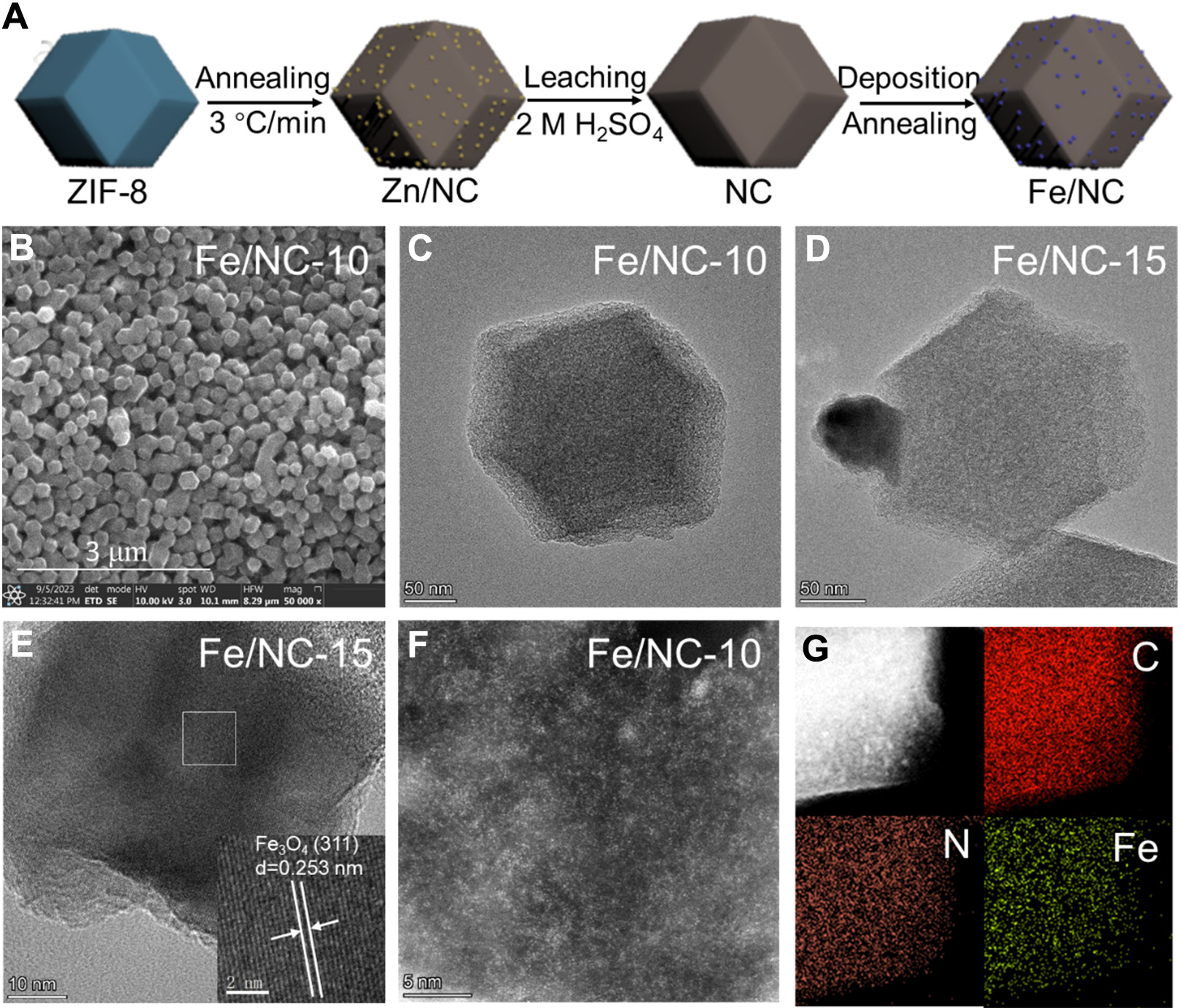
Figure 1. (A) Schematic illustration for the synthesis of Fe/NC; (B) SEM image, and (C) TEM images of Fe/NC-10; (D and E) TEM images of Fe/NC-15; (F) HAADF-STEM image of Fe/NC-10; (G) EDS mapping of Fe/NC-10, C (red), N (orange), and Fe (yellow). Three-dimensional structural models were constructed using 3ds Max 2018. SEM: Scanning electron microscopy; TEM: transmission electron microscopy; HAADF-STEM: high-angle annular dark-field scanning transmission electron microscopy; EDS: energy-dispersive spectroscopy.

