

fig1
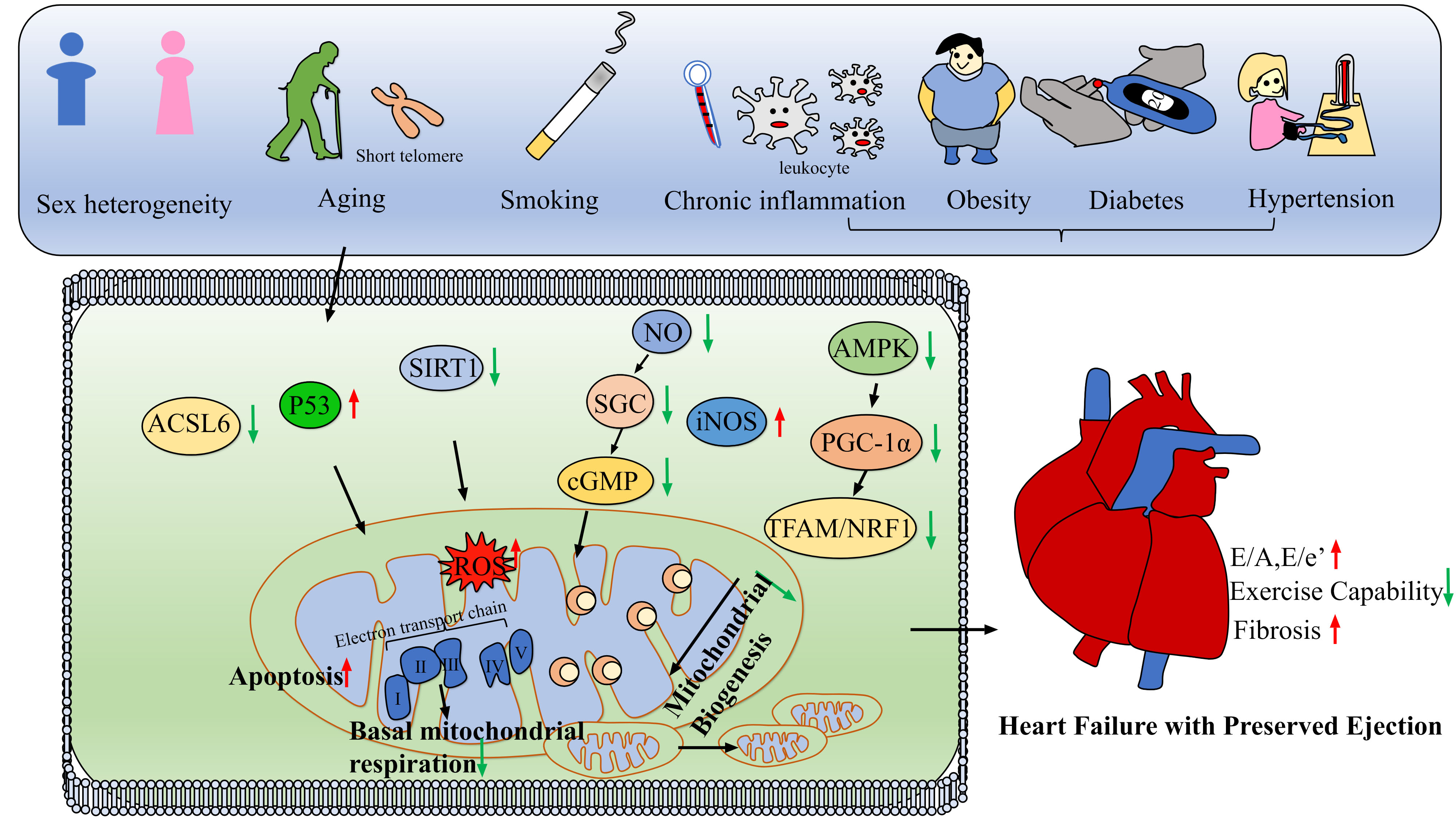
Figure 1. Pathogenesis and susceptibility of Heart Failure with Preserved Ejection Fraction (HFpEF). Schematic overview of how systemic risk factors converge on mitochondrial pathways to drive HFpEF pathogenesis. Aging, metabolic stress, inflammation/smoking, and female susceptibility disrupt key molecular hubs (p53, AMPK/PGC-1α, SIRT1, ACSL6, and NO/iNOS signaling), increase mtROS production, impair mitochondrial respiration and biogenesis, and promote apoptosis. The above mitochondrial damage is a cause of left ventricular hypertrophy and fibrosis in HFpEF, leading to diastolic dysfunction and reduced exercise capacity. In the schematic, red upward and green downward arrows indicate increased and decreased expression or activity, respectively. Black arrows indicate regulatory relationships or signaling direction. NO: Nitric oxide; mtROS: mitochondrial reactive oxygen species; iNOS: inducible nitric oxide synthase; AMPK: AMP-activated protein kinase; PGC-1α: peroxisome proliferator-activated receptor gamma coactivator 1-alpha; SIRT1: sirtuin 1; ACSL6: acyl-CoA synthetase long-chain family member 6; TFAM: mitochondrial transcription factor A; NRF1: nuclear respiratory factor 1; ROS: reactive oxygen species; SGC: soluble guanylate cyclase; cGMP: cyclic guanosine monophosphate.