

fig1
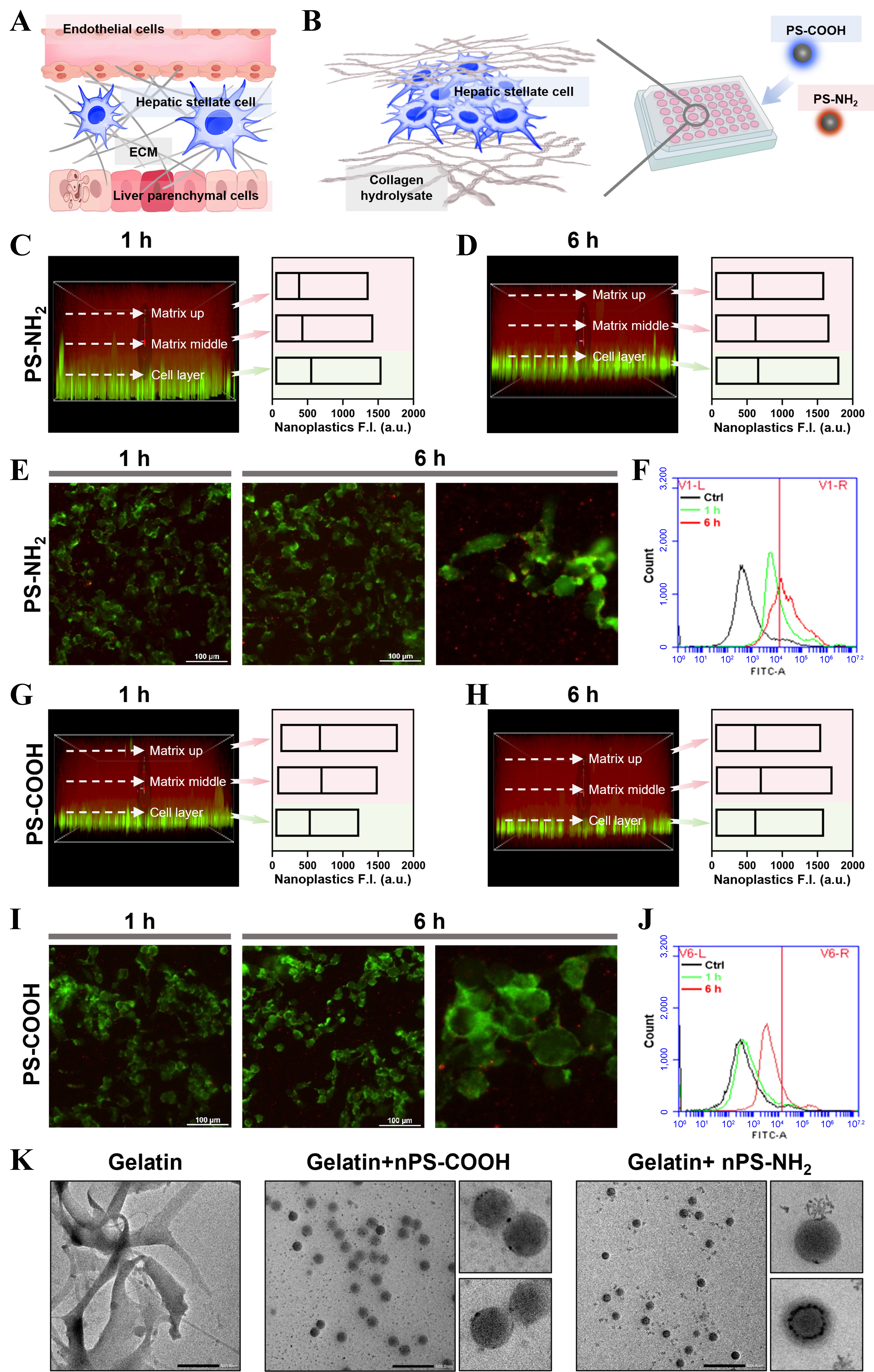
Figure 1. Differential retention and cellular uptake of positively and negatively charged nanoplastics within the 3D hepatic model. (A) Schematic illustration of the HSC microenvironment; (B) Diagram of the 3D model of the HSC treated with nanoplastics; the labeled collagen hydrolysate is gelatin; (C) Confocal z-stack images and fluorescence intensity comparison of PS-NH2 in the 3D model after 1 h exposure; (D) Confocal z-stack images and fluorescence intensity comparison of PS-NH2 after 6 h exposure; (E) Magnified fluorescence images of the cellular layer after 1 and 6 h PS-NH2 treatment; (F) Flow cytometric analysis of PS-NH2 uptake efficiency by HSCs after 1 and 6 h exposure; (G) Confocal z-stack images and fluorescence intensity comparison of PS-COOH after 1 h exposure; (H) Confocal z-stack images and fluorescence intensity comparison of PS-COOH after 6 h exposure; (I) Magnified fluorescence images of the cellular layer after 1 and 6 h PS-COOH treatment; (J) Flow cytometric analysis of PS-COOH uptake efficiency by HSCs after 1 and 6 h exposure; (K) TEM images showing gelatin adsorption on nanoplastics with different surface charges. 3D: Three-dimensional; HSC: hepatic stellate cell; PS-NH2: amino-functionalized polystyrene; PS-COOH: carboxyl-functionalized polystyrene; TEM: transmission electron microscopy; ECM: extracellular matrix.


