


fig9
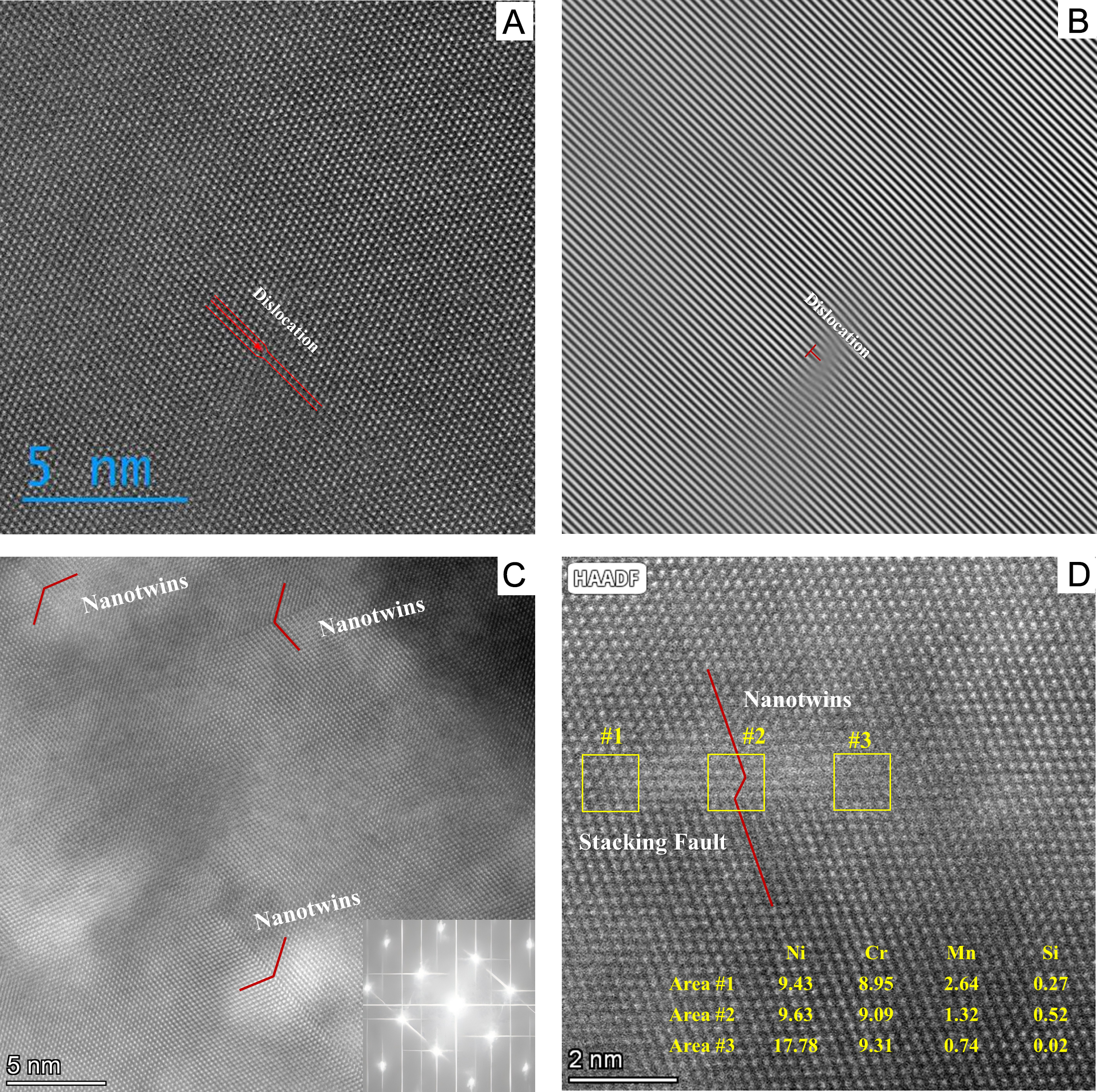
Figure 9. The atomic microstructure of FZ before and after cyclic loading at 4.2 K: (A) an STEM HAADF image of the FZ before fatigue, showing an edge dislocation, (B) IFFT of (A), (C) an STEM HAADF image of the FZ after fatigue, showing the formation of nanotwins, (D) the corresponding formation of nanotwins near the stacking faults in FZ after fatigue, with EDS results for individual elements of Ni, Cr, Mn, and Si, displaying the segregation of elements along the stacking faults and nanotwins. FZ: Fusion zone; STEM: scanning transmission electron microscopy; HAADF: high-angle annular dark-field; EDS: energy dispersive spectroscopy; IFFT: inverse fast Fourier transform.


