


fig2
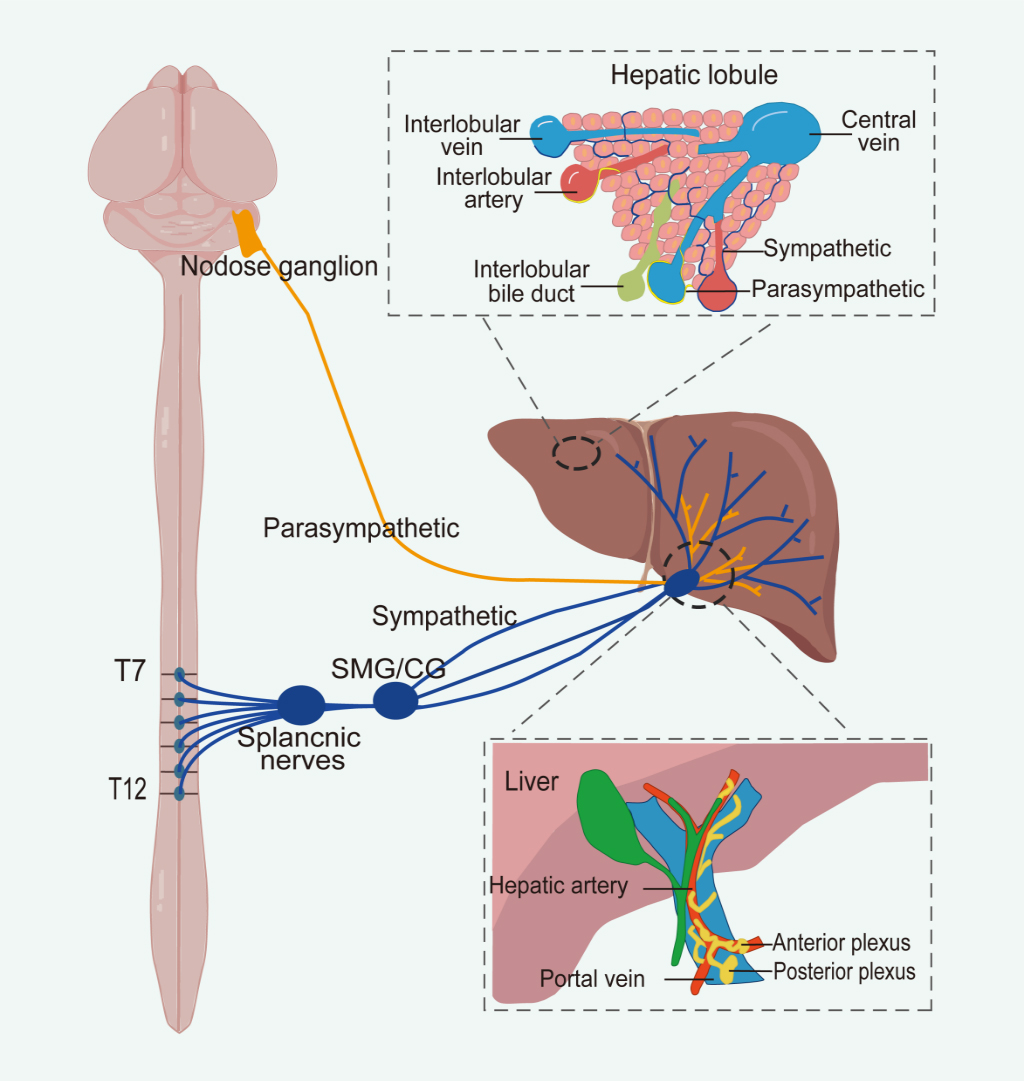
Figure 2. The sympathetic innervation of the liver arises from spinal segments T7 to T12, whereas parasympathetic innervation originates from nodose ganglion within the craniosacral regions. At the hepatic hilum, both sympathetic and parasympathetic fibers contribute to the formation of anterior and posterior autonomic plexuses. The anterior plexus surrounds the common hepatic artery, while the posterior plexus is located along the portal vein. Within the liver parenchyma, autonomic nerves are distributed around the hepatic artery, portal vein, and bile ducts. Notably, sympathetic fibers extend through the connective tissue and penetrate into the hepatic lobules, ultimately reaching the hepatocytes. Image created with Adobe Illustrator. SMG: Superior mesenteric ganglion; CG: celiac ganglion.


