

fig4
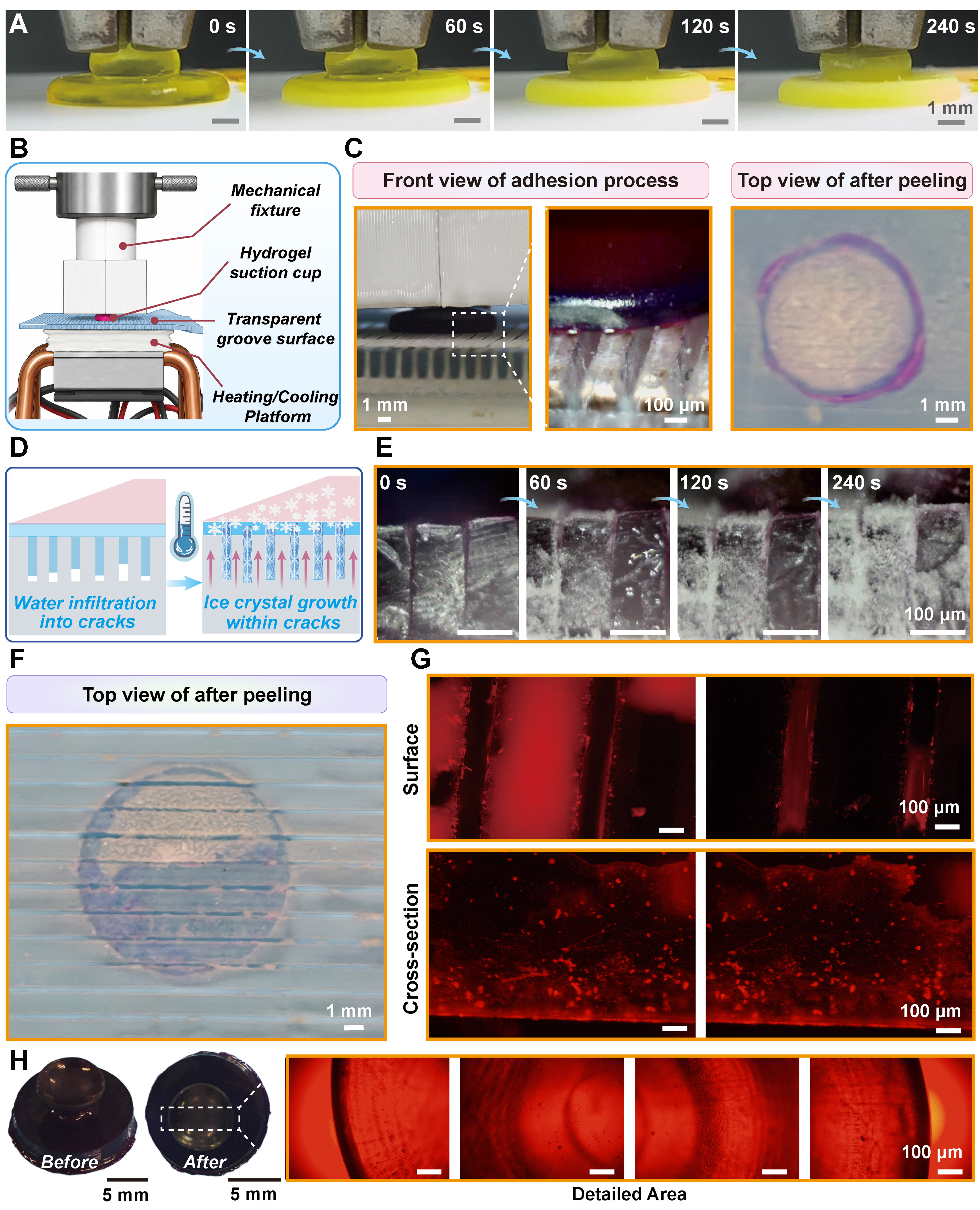
Figure 4. Evolution of the ice-activated hydrogel adhesion process. (A) Time-lapse images of freezing propagation into the hydrogel suction cup; (B) Schematic illustration of the ice-activated suction-cup adhesion testing platform; (C) Photographs showing the adhesion and peeling processes of the hydrogel suction cup; (D) Schematic illustration of ice-crystal growth and evolution in the hydrogel suction cup; (E) Photographs showing ice-crystal growth and evolution in the hydrogel suction cup; (F) Photographs of the interfacial traces after detachment; (G) Fluorescence microscopy images showing residual fluorescent traces within the interfacial grooves after detachment; (H) Photographs and fluorescence microscopy images of the hydrogel suction cup after 10 freeze–thaw adhesion cycles.