




Soft yet robust hydrogels for flexible bio-integrated devices: from mechanics properties to applications
 0
0 
Abstract
Soft hydrogels have emerged as key enabling materials for flexible bio-integrated devices owing to their tissue-like softness, high water content, and intrinsic biocompatibility. However, conventional hydrogels suffer from mechanical fragility, limited fatigue resistance, and poor environmental stability, which severely restrict their long-term performance under dynamic physiological conditions. Addressing this challenge has driven the development of combining compliant mechanics with high toughness, durability, and damage tolerance. This review provides a comprehensive overview of recent advances in robust soft hydrogels for flexible bio-integrated devices, with an emphasis on mechanics-guided design and manufacturing strategies. We first discussed molecular and network-level design principles, including chain architecture, dynamic crosslinking, double network structures, and sacrificial energy dissipation, that underpin enhanced toughness and fatigue resistance. We then examine mesoscale and microstructural engineering approaches, such as gradient architectures, anisotropic networks, and nanocomposite reinforcement, that enable mechanical programmability and environmental robustness. Fabrication and scalability strategies, including printing, patterning, and interface engineering, are subsequently reviewed to highlight pathways toward system-level integration. Finally, emerging applications in flexible electronics, electronic skin, soft robotics, and intelligent health monitoring are discussed, together with remaining challenges and future directions for translating robust soft hydrogels into reliable and manufacturable bio-integrated systems.
Keywords
INTRODUCTION
With rapid advances in materials science, electronic technology, and biomedicine, flexible bio-integrated devices have an important platform for bridging living tissues and electronic systems[1-3]. Confronted with the inherent limitations of traditional rigid electronic devices, which suffer from pronounced mechanical incompatibility with the soft, dynamic nature of biological tissues, flexible integrated devices have distinguished themselves through their excellent performances[4-7]. These devices not only exhibit exceptional conformability to biological tissues, enabling seamless mechanical and biological integration, but also unlock a plethora of cutting-edge applications, including electronic skin (e-skin)[8,9], wearable biosensors[10,11], implantable monitoring systems[12,13], and soft robotic platforms[7]. The design and development of these devices necessitate materials that not only demonstrate superior biocompatibility to ensure intimate tissue integration but also uphold long-term stability, safety, and functionality[14-16]. Consequently, the pivotal challenge propelling sustained innovation in the realm of flexible bio-integrated devices lies in achieving an optimal equilibrium between mechanical robustness and biocompatibility. Addressing this challenge is instrumental in advancing the frontiers of this burgeoning field, fostering the realization of next generation bioelectronic interfaces with unprecedented capabilities and performance.
Hydrogels, composed of hydrophilic three-dimensional polymer networks, have attracted attention as candidate materials for flexible bio-integrated devices[17,18]. Their soft, water-rich structures closely mimic the extracellular matrix, enabling excellent biocompatibility and tissue-like mechanical properties[19,20]. This unique matching minimizes foreign body response and fibrotic encapsulation, thereby supporting stable and nearly invisible interfaces with biological systems[21,22]. Beyond biocompatibility, hydrogels offer optical transparency for optogenetic and imaging applications, as well as intrinsic ionic conductivity that naturally aligns with ion-based electrophysiological signaling in living tissues. These attributes position hydrogels as highly promising flexible bio-integrated platforms for continuous health monitoring[23,24], neural stimulation[25,26], drug delivery[27,28], and even brain-computer interfaces[29,30]. However, conventional hydrogels remain far from ideal. Their mechanical fragility, reflected in low fracture toughness, poor fatigue resistance, and susceptibility to tearing, limits durability under repeated deformation in dynamic biological environments[31]. At the same time, environmental instability such as uncontrolled swelling, dehydration, and biodegradation undermines their long-term performance in vivo[32,33].These drawbacks highlight the urgent need for soft yet robust hydrogels that combine softness with mechanical resilience, where robustness is defined by high fatigue durability, enhanced fracture toughness, large elongation capacity and extended cycle life under dynamic loading.
Soft yet robust hydrogels represent a class of polymeric networks that combine high mechanical robustness with tissue-like softness, making them particularly suitable for flexible biomedical electronic applications[34-36]. Unlike conventional hydrogels that often suffer from brittleness and poor mechanical resilience, strong and tough hydrogels are characterized by their ability to withstand repeated mechanical loading without structural failure. Key indicators of their performance include fatigue durability (the capacity to resist damage under cyclic stress), fracture toughness (the energy required to propagate a crack through the material), elongation at break (the extent to which the hydrogel can stretch before rupture), and cycle life (the number of deformation cycles the hydrogel can endure while maintaining integrity). These parameters collectively determine the hydrogel’s reliability in demanding environments such as load-bearing tissue scaffolds, artificial cartilage, or long-term drug delivery systems[37,38].
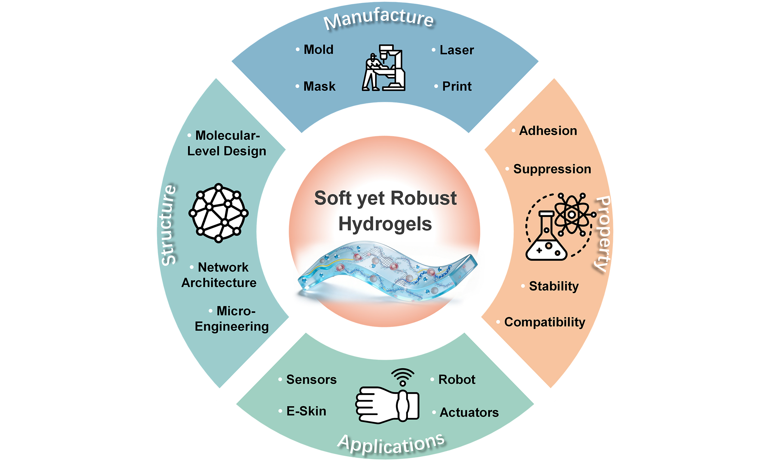
The objective of this review is to provide a comprehensive overview of recent advances in soft yet robust hydrogels for flexible bio-integrated devices, as illustrated in Figure 1. Beginning with multi-scale mechanical design strategies at the molecular, network, and macroscopic levels, we discuss how toughness, fatigue durability, and environmental stability can be enhanced through structural engineering. We then examine fabrication and scalability approaches, including advanced printing, microfabrication, and interface packaging technologies, which enable reliable integration with electronic components and biological tissues. Building on these foundations, we highlight representative applications in wearable electronics, e-skin, soft robotics, and multimodal biosensing systems, emphasizing the role of hydrogels in bridging mechanics, materials engineering, and biomedical functionality. Finally, we look ahead to emerging trends such as artificial intelligence-assisted material design, intelligent interfaces, and sustainable commercialization pathways, aiming to outline how robust hydrogel platforms can evolve into clinically viable and environmentally responsible solutions for next-generation precision medicine and health monitoring.
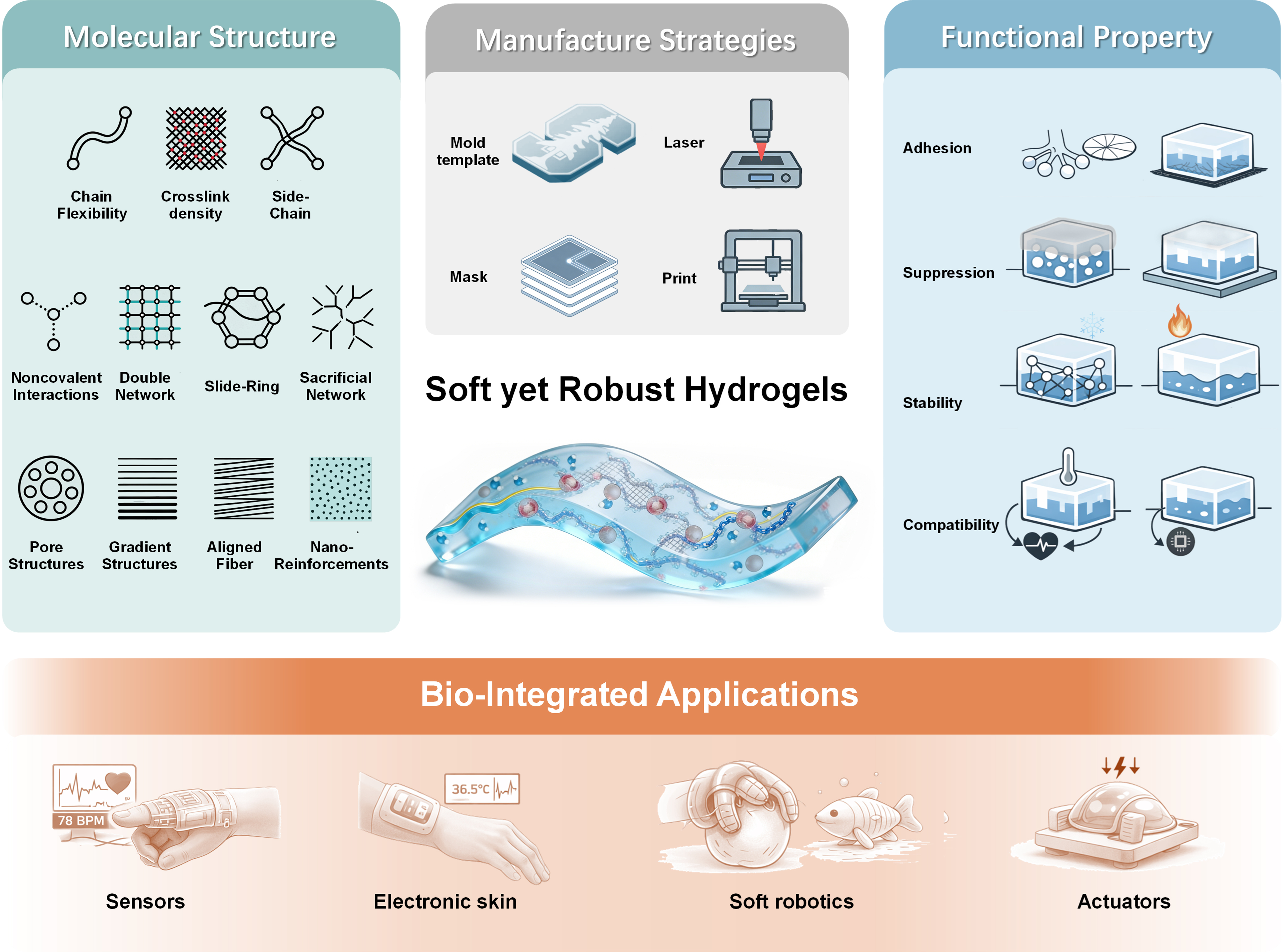
Figure 1. Overview of recent advances in soft yet robust hydrogels for flexible bio-integrated devices.
MECHANICS OF SOFT YET ROBUST HYDROGELS
Molecular/chain-level structure strategy
Molecular and chain-level structures are the key factors that determine the fundamental properties of hydrogels. By carefully tuning chain flexibility, crosslinking density, branching/side-chain architecture, and self-assembly/noncovalent interactions, the mechanical performance of hydrogels can be precisely designed
Design strategies for mechanically robust hydrogels
| Strategy | Key mechanism | Typical materials | Mechanical performance | Advantages | Ref. |
| Chain flexibility | Adjust chain mobility | Different molecular weight PVA | High stretchability, reversible large deformation | Softness, deformability, | Chen et al.[39] |
| Crosslinking density | Crosslink control | PAM + N,N′-methylenebisacrylamide | Enhanced tensile strength, hardness | Tunable strength and stability | Kim et al.[40] |
| Branching and side-chains | Entanglement and phase regulation | Hydrophobic association hydrogels | High toughness and phase stability | Functional tunability, improved stability | Pan et al.[41] |
| Noncovalent interactions | Reversible bonding | PVA–borate hydrogels | Self-healing and reversibility | Dynamic reversibility, damage recovery | Qian et al.[42] |
| Double/multiple networks | Rigid + flexible synergy | PAM + alginate DN hydrogels | High toughness and fatigue resistance | Superior toughness, load-bearing capacity | Zhou et al.[43] |
| Slide-ring structures | Stress redistribution | Slide-ring DN hydrogels | High stretchability and fatigue resistance | Stretchability, fatigue resistance | Wang et al.[44] |
| Sacrificial networks | Energy dissipation | Enzyme-cleavable peptide bond networks | High toughness and recoverability | High toughness, recoverability | He et al.[45] |
| Pore structures | Pore architecture | Granular GelMA scaffolds | High porosity and transport efficiency | Enhanced transport, tissue integration | Kedzierski et al.[46] |
| Gradient structures | Modulus gradient | PEG-based hydrogel composites | Gradient strength and modulus | Matches native tissue gradients | Eckstein et al.[47] |
| Layered/aligned fibers | Fiber orientation | CNF/MXene, CNT hydrogels | Anisotropic conductivity and toughness | Directional strength and conductivity | Geng et al.[48] |
| Nano-reinforcements | Nano reinforcement | MgFe-LDH nanosheets + chitosan/silk fibroin | High reinforcement and responsiveness | Thermoresponsive, injectable | Lv et al.[49] |
Chain flexibility
Chain flexibility reflects the ability of polymer segments to rotate and move freely in space[50]. Hydrogels with high chain flexibility allow polymer chains to adjust their conformations more easily[51], thereby imparting excellent softness and deformability [Figure 2A][40]. For example, in poly(vinyl alcohol) (PVA) hydrogels, chain flexibility can be tuned by controlling the molecular weight and degree of polymerization[39]. Wang et al. developed a PVA-based hydrogel for epidermal electronics that exhibits high stretchability, mechanical resilience, and stable sensing performance. The flexibility and mobility of PVA chains allow the hydrogel network to dynamically rearrange under external stress, allowing large and reversible deformation. Such behavior is particularly advantageous for flexible sensors subjected to frequent bending and cyclic deformation[56].
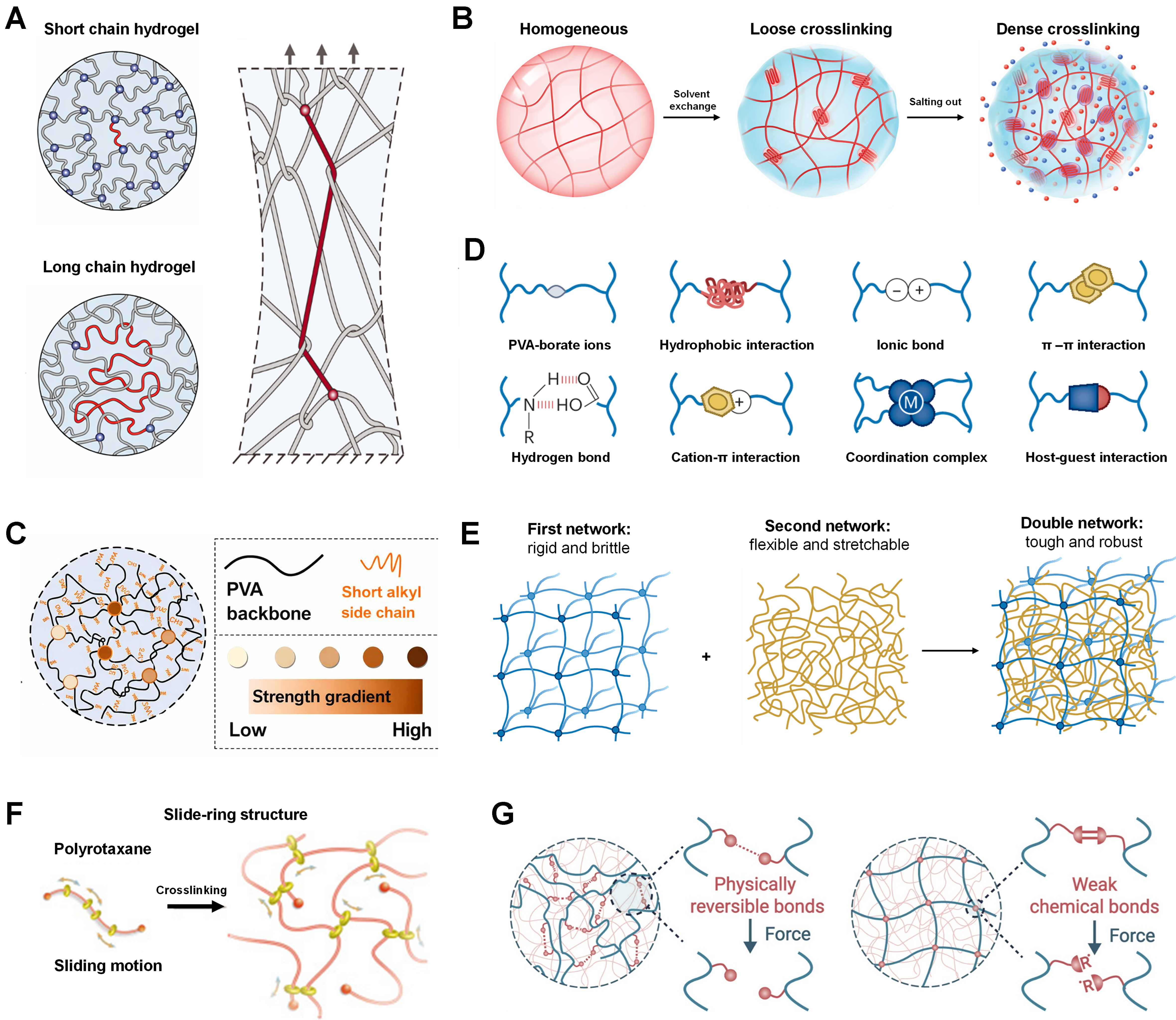
Figure 2. Molecular/chain-level structure strategy. (A) Chain flexibility tuned by molecular weight and degree of polymerization[40,52]. Copyright 2021, The American Association for the Advancement of Science; Copyright 2022, Wiley-VCH; (B) Crosslinking density[53]. Copyright 2023, Wiley-VCH; (C) Branching and side-chain structures[41]. Copyright 2021, American Chemical Society; (D) Self-assembly and noncovalent interactions; (E) Double/multiple network structures. (D and E) are reprinted with permission from Ref.[34]. Copyright 2024, Springer Nature; (F) Slide-ring and fully sliding structures[54]. Copyright 2010, Elsevier; (G) Sacrificial network design[55]. Copyright 2025, Innovation Press. PVA: Poly(vinyl alcohol).
Crosslinking density
Crosslinking density refers to the number of crosslinking points per unit volume, which plays a critical role in mechanical strength, swelling behavior, and stability [Figure 2B][53]. Increasing crosslinking density generally enhances mechanical strength by restricting chain mobility, making the hydrogel more resistant to stretching or compression[53]. However, excessive crosslinking may lead to brittleness and reduced toughness. For instance, in polyacrylamide (PAM) hydrogels, the amount of crosslinker (e.g., N, N′-methylenebisacrylamide[40]) can be precisely adjusted to control crosslinking density. Moderate increases in crosslinker content significantly improve tensile strength and hardness, but excessive amounts result in fragile hydrogels prone to fracture under stress[40].
Branching and side-chain structures
Introducing branching or side-chain architecture provides an effective strategy to regulate hydrogel mechanics and functionality. Branching increases chain entanglement, thereby enhancing mechanical strength and stability. Side chains, depending on their length, chemical nature, and spatial distribution, can impart specific functionalities[57]. For example, Pan et al. proposed a molecular design strategy to regulate phase separation in hydrophobic association hydrogels by tuning short alkyl side-chain length [Figure 2C][41]. They found that enhanced hydrophobic interactions increase the size of polymer-rich domains and shift the relaxation dynamics toward higher glass-transition temperatures. Importantly, high strength and toughness are achieved when deformation conditions match the intrinsic relaxation behavior, indicating that phase separation and glass transition jointly govern mechanical performance[41].
Self-assembly and noncovalent interactions
Self-assembly and noncovalent interactions (e.g., hydrogen bonding[58], van der Waals forces[59], π-π stacking[60]) play vital roles in hydrogel structure formation and property regulation [Figure 2D][34]. These interactions are dynamic and reversible, enabling hydrogels to undergo reversible deformation and exhibit self-healing capability. For instance, hydrogen-bonded hydrogels can reform broken bonds after damage, restoring integrity. A typical example is PVA-borate hydrogels[42], where hydroxyl groups on PVA chains interact with borate ions to form a three-dimensional network. Upon cutting or stretching, the damaged regions can self-repair through reformation of hydrogen bonds, recovering mechanical performance.
Double/multiple network structures
Double or multiple network hydrogels consist of two or more interpenetrating or interwoven networks, endowing them with superior mechanical strength and multifunctionality [Figure 2E][61]. Networks can be interconnected via chemical crosslinking or physical interactions, working synergistically to enhance toughness[62,63]. A typical double-network hydrogel combines a rigid network with a flexible one: the rigid network provides strength and resistance to stretching, while the flexible network dissipates energy and prevents catastrophic fracture[43]. This architecture yields hydrogels with high strength, flexibility, and fatigue resistance, suitable for load-bearing and wear-resistant applications.
Slide-ring and fully sliding structures
Slide-ring networks represent an advanced topological design in hydrogel mechanics [Figure 2F][54]. By introducing movable ring molecules onto polymer chains, stress can be effectively redistributed during deformation[64]. The sliding motion of rings enhances stretchability and toughness. Fully sliding structures extend this concept, allowing all network junctions to move freely, resulting in extraordinary flexibility and extensibility[54]. Wang et al. introduced slide-ring crosslinking points into a double-network (DN) hydrogel and showed that chain sliding at these movable junctions significantly enhances mechanical adaptability[44]. The hydrogel achieved reversible tensile deformation above 300%, an elastic modulus of approximately 300-500 kPa and maintained stable sensing signals after over 10,000 cyclic deformations. These results confirm that slide-ring dynamics play a critical role in mitigating stress concentration and improving fatigue resistance in flexible hydrogel systems.
Sacrificial network design
Sacrificial networks are incorporated to improve toughness and fatigue resistance [Figure 2G][55]. These networks, with relatively low mechanical strength, preferentially fracture under stress, dissipating energy and protecting the primary network[65]. By rationally designing the structure and properties of sacrificial networks, hydrogel mechanics can be finely tuned to meet diverse application requirements[66]. He et al. proposed a DN hydrogel in which the densely crosslinked sacrificial network is built from enzyme-cleavable covalent peptide bonds[45]. They demonstrated that, under force-coupled enzymatic catalysis, the sacrificial network can undergo reversible rupture and reformation, enabling recoverable energy dissipation rather than permanent damage. As a result, the hydrogel combines high toughness and reversibility[45], exhibiting fracture strains above 2,000%, a maximum toughness of 15 MJ·m-3, and a fracture energy of 9.4 kJ·m-2, while allowing mechanical recovery and self-healing after cyclic loading.
Network and microstructure regulation
Beyond molecular and chain-level structures, the network architecture and microstructural features of hydrogels also play crucial roles in determining their mechanical and functional properties. By tailoring pore structures, constructing gradient architectures, designing layered or aligned fiber structures, and incorporating nanofillers or nano-reinforcements, hydrogel performance can be further optimized.
Pore structures
Pore structures are fundamental characteristics of hydrogels, encompassing pore size, distribution, and connectivity[67]. They significantly influence stress-strain transmission and fatigue life. Larger pores facilitate transport and diffusion, enhancing swelling kinetics and drug release efficiency. However, excessively large pores may weaken mechanical strength due to thin pore walls that cannot withstand high stress. Uniform pore size distribution helps distribute stress evenly, reducing local stress concentration and improving fatigue resistance. High pore connectivity facilitates rapid mass transport throughout the hydrogel, enhancing functionality[68]. For example, in tissue engineering scaffolds, controlling pore architecture enables hydrogels to mimic the extracellular matrix by providing cell-scale void spaces and efficient nutrient transport pathways. Granular gelatin methacryloyl (GelMA) hydrogel scaffolds constructed from porous microgels exhibit hierarchical inter- and intraparticle porosity, delivering up to 170% higher void fraction and 57%-78% increase in vivo cell infiltration compared with nonporous counterparts, thereby effectively promoting cell adhesion, migration, and tissue integration. Importantly, quantitative mechanical analyses have shown that increasing pore size from ~8 to ~24 μm can redistribute stress and enhance fracture energy by approximately 2.5-fold, while simultaneously delaying fatigue crack propagation[46]. Furthermore, the polyvinyl alcohol hydrogel prepared by the freeze casting process forms a honeycomb-like porous structure [Figure 3A and B][69]. After further annealing, the crystallinity of PVA hydrogels increases by ~15%-20%, which correlates with a ~2× improvement in compressive modulus and enhanced toughness. This evidence underscores the quantitative role of pore size and connectivity in enhancing fracture toughness and fatigue resistance of hydrogel networks[69].
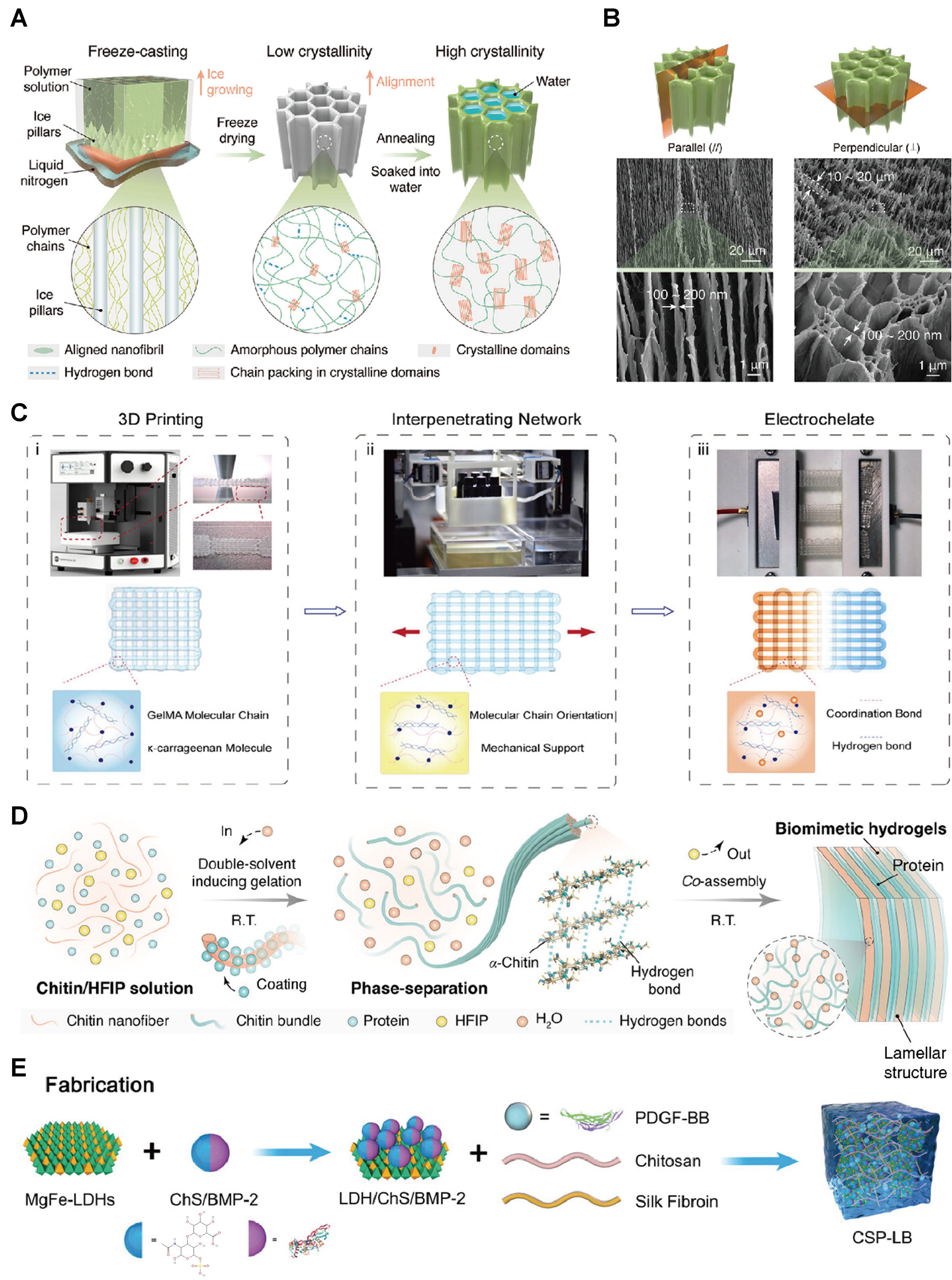
Figure 3. Design of hydrogel networks and microstructures. (A) Schematic illustration for the hydrogel fabrication by freezing a polymer solution on a copper substrate; (B) SEM images of the freeze-casted and annealed (FC-A) PVA hydrogels. (A and B) are reprinted with permission from Ref.[69]. Copyright 2021, Wiley-VCH; (C) Fabrication process: (i) 3D Printing, (ii) Mechanical Training, (iii) Electrochemical Training[74]. Copyright 2026, Wiley-VCH; (D) Preparation of biomimetic chitin-protein OfCPH-2 (BM)-hydrogel[76]. Copyright 2025, Wiley-VCH; (E) Schematic illustration of the fabrication of CSP-LB hydrogel[49]. Copyright 2023, Wiley-VCH. SEM: Scanning electron microscopy; PVA: poly(vinyl alcohol); BM: biomimetic chitin-protein OfCPH-2; CSP-LB: chitosan/silk fibroin (CS) hydrogels loaded with platelet-derived growth factor-BB (PDGF-BB) to construct a smart injectable thermo-responsive hydroge; HFIP: hexafluoroisopropanol; LDHs: layered double hydroxides; BMP-2: bone morphogenetic protein 2; PDGF-BB: platelet-derived growth factor-BB.
Gradient structures
Gradient structures are widely used to reconcile mechanical mismatches across biological interfaces[70]. Gradient structures refer to spatial variations in physical or chemical properties within hydrogels. Such architectures enable different regions to exhibit distinct performances, meeting complex application requirements, and more importantly, they redistribute stresses under cyclic loading. By gradually varying stiffness across the interface, superficial regions experience moderated strain amplitudes while deeper regions sustain lower strain levels (< 10%), thereby alleviating stress concentration, homogenizing load transfer, and delaying fatigue crack initiation during repeated deformation cycles[71]. For joint repair, hydrogels can be engineered with gradient mechanical properties so that the cartilage-side region presents a lower elastic modulus matching native cartilage (1 MPa) and mitigating local stress, while the bone-side region is substantially stiffer (5.8-6.4 MPa) to provide load-bearing support and interfacial stability[72,73]. Eckstein et al. used digital light projection/microstereolithography-printed polyethylene glycol (PEG)-based hydrogel-composite micro-truss scaffolds with sub-200 μm features and high porosity (68%-81%), the study achieved a controlled cartilage-zone gradient of 0.76-1 MPa and a 6× modulus jump into the bone layer[47]. Importantly, finite element analysis revealed that such gradients redistribute cyclic stresses: superficial regions experienced > 30% strain under compression, while deeper regions maintained < 10% strain, thereby reducing stress concentration and delaying fatigue crack initiation[47]. In addition, Yao et al. reported a preparation method termed as electrochemical training that utilizes gradient ionic coordination and molecular locking to achieve rapid assembly of disordered hydrogels [Figure 3C][74]. These hydrogels exhibit multiscale anisotropic gradient structures ranging from 5 nm to 2 cm, which not only provide spatially tuned mechanical cues but also homogenize stress transfer under repeated loading, enhancing long-term fatigue resistance.
Layered or aligned fiber structures
Layered or aligned fiber architectures introduce anisotropic mechanical, electrical, or transport properties into hydrogel systems. By controlling fiber orientation and hierarchical arrangement, hydrogels can achieve directional mechanical strength, conductivity, or optical performance[75]. For example, inspired by the layered structure of the head capsule of the Asian corn borer, a chitin hydrogel with a layered structure was developed that exhibits excellent impact resistance[76] [Figure 3D]. Conductive hydrogels can indeed be engineered by aligning conductive micro/nanofibers [e.g., cellulose nanofiber (CNF)/MXene, carbon nanotubes (CNTs)] into an ordered fiber network, which produces direction-dependent electrical pathways[76]. Representative systems show that the conductivity parallel to the fiber alignment markedly exceeds the perpendicular direction: for example, an anisotropic CNF/MXene DN hydrogel reaches
Nanofillers and nano-reinforcements
Incorporating nanofillers or nano-reinforcements provides an effective route to enhance hydrogel mechanical and functional performance. Nanomaterials possess unique physical and chemical properties, such as high surface area, superior mechanical strength, and distinctive optical or electrical characteristics[77]. Uniform dispersion of nanomaterials (e.g., CNTs[78], nanofibers[79], nanosheets[49]) within hydrogel matrices can significantly improve mechanical strength, conductivity, thermal stability, and bioactivity. Lv et al. proposed incorporating MgFe-layered double hydroxide (LDH) nanosheets into a chitosan/silk fibroin hydrogel to reinforce the polymer network and tune its gelation and mechanical properties [Figure 3E][49]. They showed that the nanosheets act as physical crosslinking and load-bearing units, significantly shortening gelation time (from 300 to 146 s), lowering the sol-gel transition temperature (37.4 to 32.7 °C), and enhancing the storage and compressive moduli by 12-fold and 10-fold at optimal LDH content (0.1 wt%). Based on these nanosheet-reinforced mechanical properties, the hydrogel was further demonstrated as an injectable thermos-responsive platform for effective bone regeneration.
Fatigue performance and environmental stability
In emerging applications such as flexible electronics, soft actuators, biomimetic materials, and wearable devices, the fatigue performance and environmental stability of materials are critical determinants of long-term service reliability. Soft materials are frequently exposed to complex environmental conditions, including variations in humidity, temperature, and ionic strength, while simultaneously undergoing repeated mechanical deformation such as stretching, compression, or bending. Under these conditions, cyclic loading behavior, moisture exchange, and structural relaxation processes collectively determine the functional lifespan and structural integrity of hydrogel systems. This section reviews recent advances in three key aspects: (i) fatigue behavior under cyclic loading across different humidity, temperature, and salt environments; (ii) performance degradation induced by moisture loss, evaporation, and dry-wet cycling; and (iii) long-term reliability in maintaining deformation recovery and mechanical strength.
Fatigue behavior under cyclic loading
Materials used in practical applications are frequently subjected to repeated mechanical deformation, making resistance to mechanical fatigue a key design criterion[69]. Environmental conditions have been widely shown to exert a strong influence on material performance during cyclic loading. In high humidity environments, water molecules can penetrate polymer networks and act as plasticizers, reducing modulus while enhancing chain mobility[80]. Although this effect may temporarily improve flexibility, prolonged exposure often disrupts crosslinking structures and accelerates crack propagation. For example, hydrogels typically exhibit good stretch recovery behavior under humid conditions but may suffer from localized dehydration and pore collapse during prolonged compressive cycling, resulting in increased energy dissipation and accumulation of residual strain[81].
Elevated temperatures intensify thermal motion within polymer networks, potentially leading to chain disentanglement, crosslink cleavage, or phase separation, thereby reducing fatigue life[82]. In contrast, low temperatures can embrittle soft materials by suppressing energy dissipation mechanisms, increasing susceptibility to brittle fracture[83]. High salt environments can also significantly influence hydrogel mechanics by screening electrostatic interactions or modifying ionic crosslinking within polymer networks[84]. However, such salt-induced stiffening is often accompanied by reduced elasticity and flexibility of the hydrogel network.
Fatigue-resistant mechanism
The core of material fatigue-resistant design is to construct a hierarchical protection system integrating crack suppression, energy dissipation and damage self-repair[85,86]. While all fatigue-resistant designs aim to restrain crack expansion and reduce cumulative cyclic damage, three mainstream design strategies differ substantially in structural design principles, working mechanisms and fatigue-resistant performances, as summarized in Table 2.
Comparison of typical fatigue-resistant design strategies for functional soft materials
| Design strategy | Core design principle | Main fatigue-resistant mechanism | Key performance characteristics | Limitations |
| Crystalline domain regulation[87] | Optimize grain size, distribution, and crystallinity; exert synergistic effect of crystalline and amorphous regions | Block crack propagation, dissipate strain energy, relieve stress concentration | High initial modulus and strength; relatively low fatigue threshold; fast crack propagation rate under large strain | Poor ductility, unsuitable for high-strain long-cycle fatigue service |
| Soft network topology modulation[89] | Construct interpenetrating, sliding, dynamic reversible and high-functional cross-linked networks | Realize hierarchical energy dissipation, uniform stress distribution and microdamage self-repair | Excellent large deformation adaptability, slow crack growth rate, superior long-cycle fatigue stability | Weak high-temperature resistance, easy mechanical attenuation under extreme external load |
| Multiphase composite reinforcement[90] | Build stable filler-matrix interface, optimize interfacial bonding and gradient structure | Reinforcing phase bridging effect, efficient interfacial load transfer, interfacial energy dissipation | Adjustable fatigue performance, improved environmental adaptability, balanced mechanical properties | Prone to interfacial debonding; complex preparation process, difficult precise regulation |
Crystalline domain modulation represents a typical fatigue improvement strategy applicable to crystalline substances and crystalline polymer composites[86]. Its core design principle focuses on precise regulation of grain size, spatial distribution, and overall crystallinity, and exploits the synergistic interaction between crystalline and amorphous phases to optimize internal stress distribution. This structural optimization strengthens intrinsic crack resistance, realizes efficient strain energy dissipation, and relieves local stress concentration. For instance, in high-molecular-weight PVA hydrogels, cracks grow through the combination of chain sliding and chain breakage. The crystalline domains reduce stress near the crack tip, and the threshold increases with the increase in crystallinity[87]. At 18.9 wt% crystallinity, the threshold can exceed 1,000 J/m2. It should be noted that brittleness dominates and fatigue resistance deteriorates beyond ~70% crystallinity[87]. Such rigid structural regulation is limited by low ductility, which usually results in relatively low fatigue threshold and rapid crack propagation rate under large cyclic deformation, restricting its application in high-strain fatigue scenarios.
In sharp contrast, internal network topology engineering is the dominant design route for flexible soft materials to achieve excellent fatigue resistance[88]. Distinct from rigid crystalline reinforcement, this strategy relies on customized network architectures including interpenetrating networks, sliding crosslinking networks, dynamically reversible crosslinked networks, and high-functionality crosslinked networks. Its unique design logic lies in realizing multi-stage energy dissipation, homogeneous stress redistribution and spontaneous microdamage rehabilitation through flexible chain motion and reversible bond dynamics. For example, a double-network elastomer can achieve fatigue thresholds as high as 500 J/m2[89], but at the cost of substantially lower initial modulus (often < 1 MPa vs. > 10 MPa for crystalline materials). Furthermore, dynamic reversible cross-links further enable micro-damage self-repair under cyclic loading, a capability absent in purely crystalline designs. Benefiting from such flexible structural characteristics, network-regulated soft materials possess outstanding long-cycle fatigue tolerance and slow crack propagation speed, yet their fatigue stability is easily deteriorated under high-temperature and high-load service conditions.
Different from the above two strategies, the fatigue-resistant performance of multi-phase composite materials is dominated by reinforcing phase bridging behavior and interfacial load transfer efficiency[90]. Its design philosophy centers on constructing stable filler-matrix interfacial interaction: reinforcing phases across crack gaps generate crack closure force and consume interfacial fracture energy to slow down crack growth. Further interfacial modification strategies including bonding strength optimization, gradient interfacial construction and high-energy phase introduction can effectively block crack extension paths and elevate the critical fatigue threshold of composites. In the optimized system, the fatigue threshold has exceeded 1,000 J/m2[91]. However, unreasonable interfacial matching easily causes interfacial debonding failure, which becomes the main factor restricting the further promotion of comprehensive fatigue performance.
In summary, the three strategies show obvious differences in key fatigue-related quantitative indicators: crystalline regulation achieves high static mechanical strength but poor cyclic fatigue durability; network topology design balances large deformation adaptability and long-cycle stability; composite reinforcement realizes performance complementarity at the expense of increased structural complexity. Recently, emerging advanced material systems integrate the merits of the above design concepts to obtain optimized fatigue resistance under complex service environments, such as dual-network hydrogels, nanofilled elastomers and bionic layered structural materials. Specifically, bidirectional freeze-casting induced layered microstructure provides a feasible route to fabricate isotropic fatigue-resistant materials[76,92,93]. Hydrogels with ordered 2D layered structures and finely tuned nanocrystalline domains achieve greatly improved comprehensive mechanical properties[94]. The introduction of rigid nanofillers such as cellulose nanocrystals and MXene can further suppress crack initiation and propagation, endowing materials with reliable fatigue-resistant capability in humid service environments [Figure 4A][95,96]. Moreover, gradient crosslinking design optimizes internal stress field distribution and alleviates local stress accumulation, which is an effective auxiliary means to further enhance the cyclic structural resilience of fatigue-resistant materials.
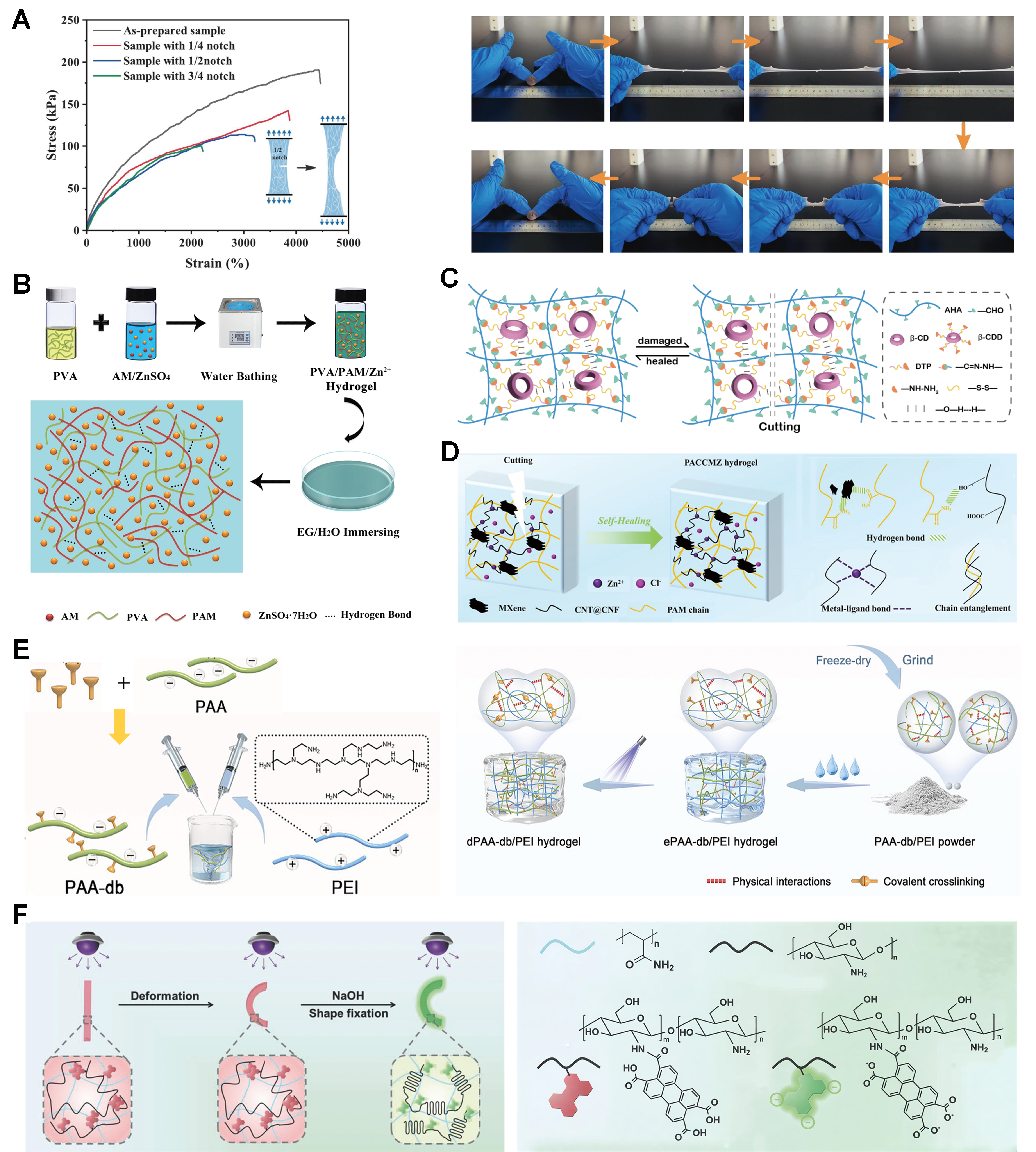
Figure 4. Fatigue performance and environmental stability of hydrogels. (A) Photos of the stretched hydrogel with a notch and tensile stress-strain curves[96]. Copyright 2025, Elsevier; (B) Schematic illustration on the synthesis and structure of PVA/PAM/Zn/EG (PPZE) hydrogel[98]. Copyright 2022, The American Association for the Advancement of Science; (C) Self-healing mechanism of β-cyclodextrin/hyaluronic acid (ACD) hydrogel[104]. Copyright 2025, Elsevier; (D) Self-healing mechanism diagram of PACCMZ hydrogel. Self-healing mechanism[103]. Copyright 2025, Elsevier; (E) Schematic diagram for the preparation of negatively-charged double bond-functionalized polyacrylic acid and positively-charged polyethyleneimine powder (PAA-db/PEI powder)[112]. Copyright 2025, Wiley-VCH; (F) The mechanisms of the shape memory hydrogels with simultaneously switchable fluorescence[113]; Copyright 2018, Wiley-VCH. PVA: Poly(vinyl alcohol); PAM: polyacrylamide; EG: ethylene glycol; PACCMZ: polyacrylamide/carbon nanotube@cellulose/MXene/Zn2+; AM: acrylamide; AHA: aldehyde-modified hyaluronic acid; DTP: 3,3′-dithiobis (propionyl hydrazide); CNT: carbon nanotube; CNF: cellulose nanofiber; PAA: polyacrylic acid; PEI: polyethyleneimine.
Moisture-induced performance degradation
Moisture exchange is a central factor governing the environmental stability of soft materials, particularly hydrophilic systems such as hydrogels and hygroscopic polymers. Repeated drying and wetting cycles often induce irreversible structural damage. Under low humidity conditions, water evaporation leads to volumetric shrinkage, pore collapse, and network densification, resulting in increased Young modulus at the expense of ductility[97]. This drying-induced contraction is commonly accompanied by interfacial delamination and surface cracking, severely limiting material reusability.
More critically, repeated dry-wet cycling causes cumulative structural degradation. Although rehydration may partially restore volume, incomplete network reconstruction frequently leads to anisotropic swelling, microcrack propagation, and blockage of ion transport pathways. Rapid evaporation can further generate capillary forces that induce macroscopic wrinkling and delamination, undermining structural integrity. Strategies such as incorporating alcohol have been reported to improve water retention in hydrogels [Figure 4B][98]. Nevertheless, complete prevention of water loss remains challenging.
To address these issues, several approaches have been proposed, including construction of amphiphilic networks using block copolymers or organic inorganic hybrids to regulate water diffusion kinetics[99,100], integration of self-healing functionalities through dynamic covalent bonds (e.g., imine or boronate ester linkages) or noncovalent interactions such as hydrogen bonding and metal coordination. to enable post damage repair [Figure 4C and D][101-104], and application of encapsulation strategies such as polydimethylsiloxane (PDMS) coatings or microencapsulation to physically shield active layers from environmental exposure[105,106].
Long-term mechanical reliability
Beyond short term cyclic loading, materials deployed in practical systems must maintain mechanical performance under prolonged static or quasi-static deformation. Most polymer materials, however, exhibit creep and stress relaxation[107], manifested as progressive deformation under constant load or gradual decay of stress over time, posing challenges for applications requiring dimensional stability or long-term sealing.
Environmental factors further exacerbate these phenomena. Elevated temperatures accelerate molecular chain slippage and enhance stress relaxation rates[108], while humidity fluctuations alter free volume and dielectric properties, disrupting internal energy dissipation mechanisms[109]. To improve long term reliability, modern material design increasingly emphasizes multiscale stable network architecture. Crystallizable elastomers, for example, employ reversible crystalline domains as physical crosslinks to preserve modulus even at elevated temperatures[110]. Shape memory polymers provide the ability to recover original shape upon external stimulation, extending effective service life[111]. In addition, environmentally responsive crosslinking modulation, such as photo triggered or pH sensitive networks, enables mechanical reinforcement and opens new opportunities for adaptive material systems [Figure 4E and F][112,113].
Conductive mechanisms and electrical design of soft hydrogels
Conductive hydrogels have attracted considerable attention due to their ability to transport ions or electrons, making them a rapidly emerging focus in materials science and biomedical research. Based on their conduction mechanisms, conductive hydrogels can be categorized into three types: ionic conductive hydrogels, electronically conductive hydrogels, and hybrid conductive hydrogels. This section provides a comprehensive overview of their conductive behaviors, with emphasis on the underlying mechanisms, key influencing factors, and potential applications. The discussion aims to offer theoretical insights and practical guidance for the design and utilization of conductive hydrogels in diverse fields.
The conductive strategies outlined in the previous section effectively overcome the challenge of electrical signal transmission, thereby establishing a foundation for developing “electronic-biological” interfaces. However, for devices to progress beyond functioning as passive signal conduits and advance into intelligent systems that actively adapt to dynamic physiological environments while performing specific functions, it is crucial to endow materials with controllable, multimodal responsiveness to environmental stimuli[114-118]. Hydrogels exhibiting such responsiveness can transduce external physical or chemical stimuli (such as temperature, pH, light, electricity, magnetism, or specific biomolecules) into observable and functional output signals, including changes in volume, shape, color, or optoelectronic properties, thus enabling real-time feedback on physiological states and intelligent simulation of environmental interactions[119-121]. Among these responses, volume change represents the most fundamental form, which can be strategically guided toward complex deformations through spatial structural design[122,123]. Furthermore, coupling periodic structures with volume variations facilitates visualized color feedback, while the integration of light-harvesting moieties with charge transport networks unlocks pathways for efficient optoelectronic conversion. This section systematically elaborates on how hydrogels, via multiscale design, integrate diverse responsive mechanisms and sensing functions to achieve a transition from simple conduction to complex behaviors in flexible bio-integrated devices.
Ionic conductive hydrogels
Ionic conductive hydrogels conduct electricity through the directional migration of mobile ions within their hydrated polymer networks. Owing to their high water content, ions such as H+, OH-, Na+, and Cl- are readily generated by water dissociation or polymer ionization[124]. Under an external electric field, these ions migrate along concentration or potential gradients, producing ionic current. For example, in sodium polyacrylate-based hydrogels, partial dissociation of sodium carboxylate groups releases Na+, and carboxylate counterions, which act as charge carriers within the three-dimensional network[125]. Ionic conductivity is primarily governed by ion concentration, network architecture, and temperature. Higher ion density increases the number of charge carriers, while a loose and porous network provides low-resistance pathways for ion diffusion. Elevated temperature enhances ionic mobility by accelerating segmental motion and diffusion, although excessive heating may destabilize the hydrated structure and compromise conductivity[126]. Importantly, because ionic transport depends on hydrated channels, microcracks or dehydration induced by mechanical fatigue can interrupt ion pathways, linking conductivity decay directly to structural durability.
Electronically conductive hydrogels
Electronically conductive hydrogels rely on electron transport through embedded conductive phases within an otherwise insulating polymer matrix. This is commonly achieved by introducing conductive fillers such as CNTs[78], graphene[127], or metallic nanoparticles[128], that form percolated networks or continuous conductive pathways. When a sufficient filler concentration is reached, electrons can hop between adjacent fillers or travel along interconnected networks under an electric field. For instance, graphene sheets dispersed in a PVA hydrogel can overlap to form conductive channels, enabling efficient electron transport[127]. The conductivity of electronically conductive hydrogels depends strongly on filler type, loading level, dispersion quality, and interfacial interactions with the polymer network. Well-dispersed fillers with strong interfacial bonding facilitate continuous conductive pathways, whereas aggregation or poor compatibility disrupts electron transport. In addition, the hydrogel’s network flexibility and porosity influence the stability of conductive networks during deformation. Large strains or repeated fatigue cycles may fracture conductive bridges, showing how mechanical resilience and electrical performance are inherently coupled.
Hybrid conductive hydrogels
Hybrid conductive hydrogels combine ionic and electronic conduction mechanisms by simultaneously incorporating mobile ions and electronically conductive components within a single hydrogel matrix. In such systems, ionic species migrate through hydrated polymer domains, while electrons are transported through interconnected conductive networks[129]. For example, sodium polyacrylate can provide ionic conduction via dissociated Na+ ions, whereas polyaniline (PANI) embedded in the same matrix enables electronic conduction through percolated networks[130]. The coexistence of these two charge transport pathways results in synergistic conductive behavior. The relative contribution of ionic and electronic conduction can be tuned by adjusting the composition ratio, dispersion state, and interactions between the ionic and electronic components. Environmental factors such as temperature and humidity further modulate performance by influencing ion mobility and the integrity of electronic networks. While dual pathways provide redundancy, mechanical damage such as crack propagation or network collapse can simultaneously impair both ionic and electronic channels, underscoring the need for architectures that balance conductivity with fatigue resistance[131].
In summary, fatigue performance and environmental stability of soft materials in humid, thermal, and saline environments are governed by the coupled effects of moisture migration, ionic interactions, and thermally activated processes. Although substantial progress has been achieved in enhancing fatigue resistance and environmental adaptability, several challenges remain. These include achieving simultaneous optimization of conductivity, flexibility, and durability under high humidity and high salt conditions, establishing standardized metrics for quantitative evaluation of structural reversibility during dry-wet cycling, and developing in situ monitoring techniques capable of tracking aging mechanisms during long term operation.
Future research should focus on intelligent and adaptive material systems supported by in situ characterization tools such as Raman spectroscopy and atomic force microscope-infrared (AFM IR) spectroscopy to elucidate microstructural evolution during service[132,133]. Integration of machine learning models to predict lifetime performance based on coupled environmental and mechanical inputs will further enable rational design and engineering scale deployment of durable, high-performance soft functional materials.
FABRICATION AND SCALABILITY STRATEGIES
Assembly strategies towards soft hydrogels
In view of their high-water content and mechanically compliant properties, hydrogels are incompatible with the high-temperature and high-vacuum processing methods typically employed in the fabrication of conventional rigid electronics[134-136]. As a result, innovative manufacturing techniques have been created specifically for hydrogel-based bioelectronics. This section reviews these methods, such as solvent casting, layer-by-layer (LbL) assembly, photolithography, laser-assisted patterning, and screen printing, 3D printing technique [Table 3].
Fabrication strategies for robust hydrogels used in flexible bio-integrated devices
| Strategies | Principle | Achievable structures | Advantages | Representative applications | Ref. |
| Free-radical polymerization | Free radical-initiated monomer crosslinking into 3D networks | Bulk, thin films and micro-patterned | Simple, fast, controllable and scalable | Flexible electrodes and tissue scaffolds | [137] |
| Click chemistry polymerization | Specific click reactions form covalent networks | Precise, core-shell and functionalized | Mild, specific and biocompatible | Injectable adhesives and glucose sensors | [138] |
| Double-network fabrication | Two interpenetrating networks dissipate energy | Tough bulk and flexible films | Tough, stretchable and fatigue-resistant | Wearable sensors and artificial skin | [139] |
| Electrochemical polymerization | Monomer polymerization on electrodes via electric field | Electrode films and conductive composites | Good adhesion, controllable and conductive | Neural and EMG sensors | [140] |
| 3D printing | LbL precursor deposition for custom 3D structures | Custom porous and personalized devices | Precise, customizable and multi-material | Personalized sensors and cartilage scaffolds | [141] |
| Ionic crosslinking | Electrostatic interactions form physical networks | Injectable, bulk and tunable films | Mild, fast, non-toxic and injectable | Wound dressings and drug carriers | [142] |
Solvent casting is a commonly used molding process. After dissolving or dispersing the base materials and functional additives in a solvent, the mixture is cast into a mold and the solvent is permitted to evaporate, leading to hydrogel formation[143,144]. For direct fabrication of conductive hydrogels bioelectronics using templating, hydrogel-based precursors are poured into a specially designed mold, ensuring precise alignment with the intended patterns. The hydrogel precursor is then cured in place through methods such as chemical crosslinking, ultraviolet (UV) polymerization, air drying, or other curing techniques, which solidify the hydrogel network. This technique is low-cost and ideal for the large-scale preparation of simple hydrogel architectures, which also allows for distribution of nanoparticles or other functional elements throughout the hydrogel, while the evaporation process helps control both the material’s microscopic structure and overall performance. For example, Chen et al. in Figure 5A introduced a one-step method that uses hydronium ion-driven dissociation along with chemical cross-linking to quickly dissolve and modify cellulose, resulting in highly organized and durable cellulose films[145]. In their process, an organic acid creates a strong driving force for H+ release, which, together with H2SO4, triggers both the dissolution and derivatization of cellulose. Similarly, Han et al. in Figure 5B developed a conductive, soft, and transparent hydrogel by integrating biocompatible PVA and poly(vinylpyrrolidone) (PVP) chains[146]. This hydrogel features an extremely low modulus, excellent resilience, and high transparency. Additionally, polydopamine nanoparticles are embedded within the hydrogel structures, greatly improving their adhesive strength and electrical conductivity, all while maintaining superior transparency. Overall, solvent casting remains one of the most widely used techniques for making hydrogel electronics, offering extensive material compatibility with few limitations on material types. However, constraints related to mold size and difficulties in separating the finished product can limit large-scale manufacturing and high-precision production of hydrogel bioelectronics[147,148]. However, despite its simplicity and material versatility, solvent casting is limited in achieving high-resolution patterning and precise structural control, which restricts its application in miniaturized and high-density bioelectronic systems.
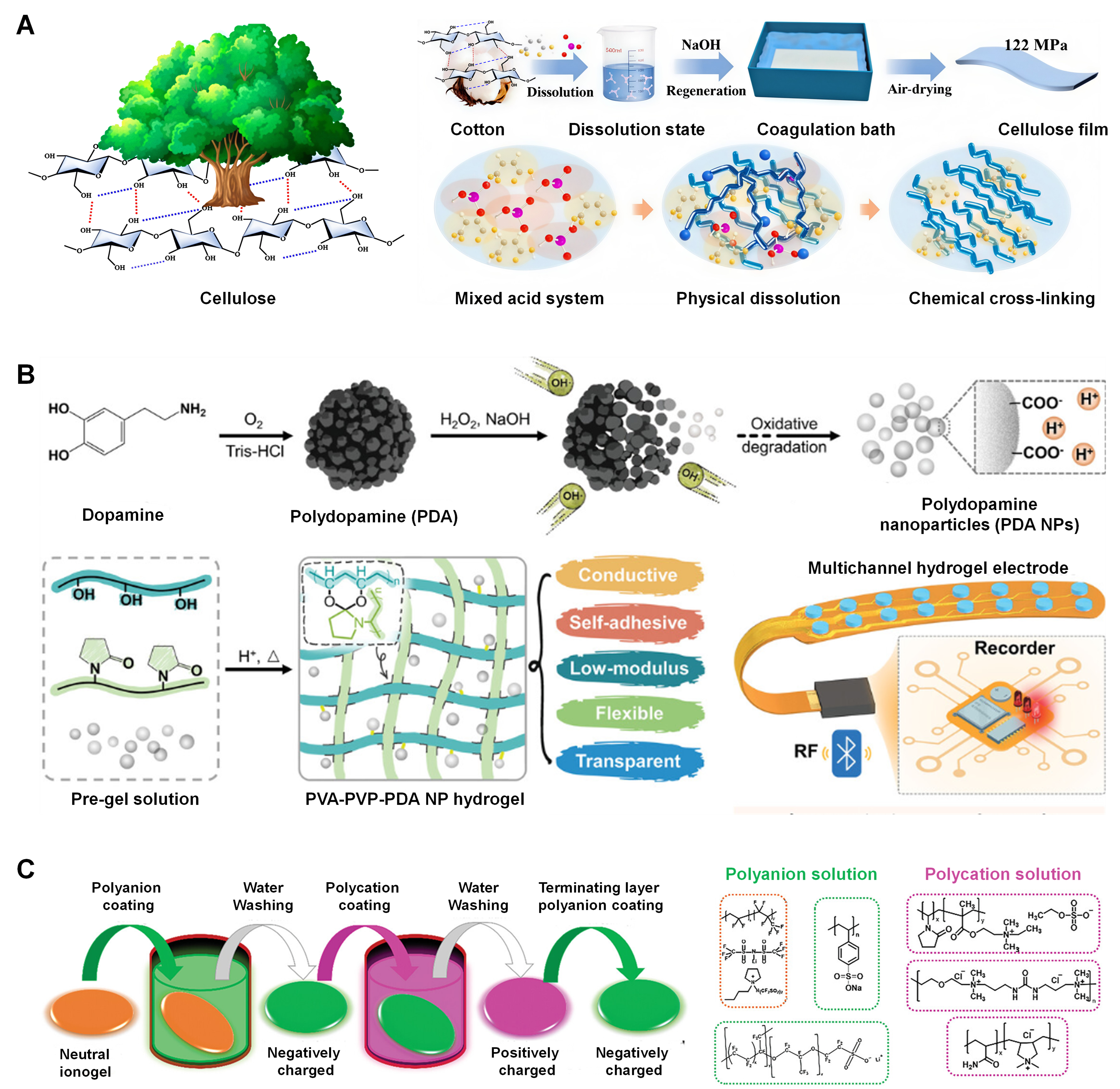
Figure 5. (A) One-pot strategy for dissolving cellulose hydrogels[145]. Copyright 2024, American Chemical Society; (B) Hydrogels from PVA-PVP-PDA NPs materials[146]. Copyright 2023, Wiley-VCH; (C) The ionogel via the LbL assembly approach[155]; Copyright 2025, Wiley-VCH. PVA: Poly(vinyl alcohol); PVP: poly(vinylpyrrolidone); PDA: polydopamine; NPs: nanoparticles; LbL: layer-by-layer; RF: radio frequency.
LbL assembly enables highly precise control over the composition, thickness, and interfacial characteristics of materials, particularly at the nanoscale[149,150]. In this approach, functional components are deposited onto a substrate sequentially, one layer at a time, with each layer stabilized through interactions such as hydrogen bonding or covalent bonding[151,152]. Electrostatic forces are crucial for maintaining the stability of these multilayer structures and allow for fine-tuning of the coating’s thickness and surface features. With LbL assembly, it’s possible to create bilayer or multilayer structures on a substrate, each with unique functionalities[153,154]. For instance, Thapaliya et al. in Figure 5C developed polyelectrolyte membranes with high charge density by alternately layering polyions with specially designed functional groups onto a neutral ionogel membrane, leveraging the ionogel’s inherent ionic nature for easy interfacial modification[155]. The LbL technique enables precise, molecular-level control over the membrane’s structure, allowing for the integration of functional groups specifically suited for lithium metal battery applications. As a result, these LbL-assembled polyelectrolytes demonstrate enhanced mechanical strength and electrochemical stability, making them ideal for use in high-voltage environments[156].
Creating hydrogel bioelectronics with photolithography involves applying a hydrogel precursor onto a substrate and then exposing it to UV light through a photomask to form a specific pattern[157,158]. Once exposed, the photoresist layer is removed, revealing the patterned hydrogel structures underneath. However, because hydrogels contain a lot of water and tend to swell when they come into contact with the precursor solution, overexposure between neighboring features often restricts the resolution to around 100 µm. As a result, traditional photolithography has difficulty achieving high-precision and direct patterning of conductive hydrogels[158]. For instance, Liu et al. in Figure 6A and B created a hydrogel-based electrode with a resolution as fine as 5 µm by applying standard photolithographic patterning to an ion gel [poly(3,4-ethylenedioxythiophene):poly(styrenesulfonate) (PEDOT:PSS)-ionic liquid (IL) ion gel], which was then converted into a micropatterned hydrogel structure[159]. They effectively solved the problem of uncontrolled swelling in the micropatterned hydrogels by using a solvent exchange process. After curing the precursor with UV light, the resulting patterns showed excellent elasticity, flexibility, and chemical stability in most solvents. Compared to other fabrication techniques, photolithography enables the production of hydrogel bioelectronics with extremely detailed features, reaching down to just a few micrometers[160,161]. However, this method is typically limited to making two-dimensional planar patterns, and any changes to the electrode design require a new photomask, which can lower efficiency and raise costs[160,161]. Additionally, the light sources used in photolithography may damage the hydrogel network, especially in systems that include living components like enzymes or bacteria.
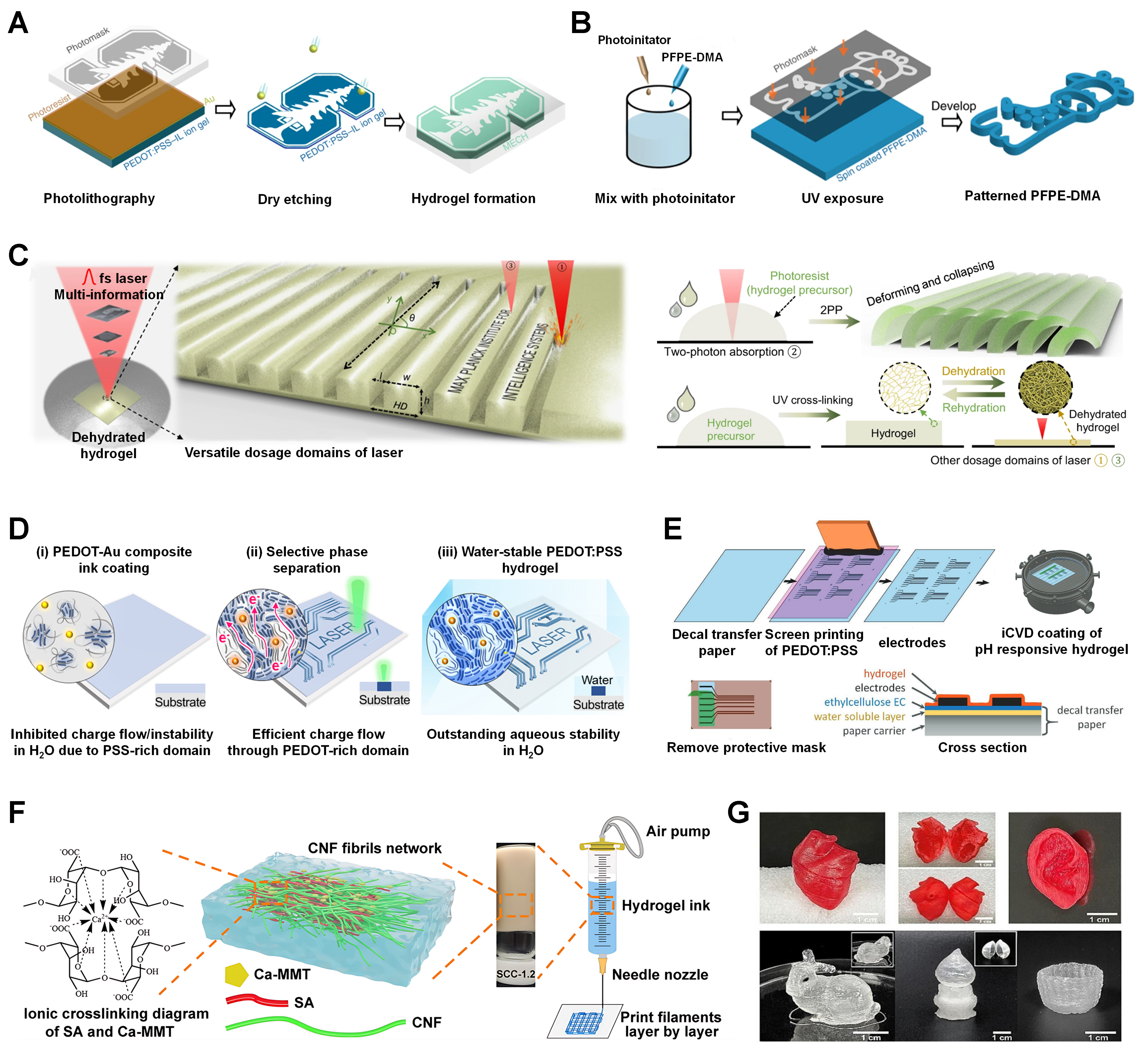
Figure 6. (A) Lithographically assisted method for hydrogels. cellulose hydrogels; (B) Stepwise depiction of hydrogel lithography. (A and B) are reprinted with permission from Ref.[159]. Copyright 2019, Springer Nature; (C) Laser technology for the dehydrated hydrogel film[160]. Copyright 2023, Springer Nature; (D) Laser-induced method for conducting polymer[163]. Copyright 2022, The American Association for the Advancement of Science; (E) A transferable temporary tattoo[169]. Copyright 2022, Wiley-VCH; (F) Direct-ink-writable hydrogel systems[174]. Copyright 2024, Elsevier; (G) 3D-printing regenerated cellulose hydrogels[175]. Copyright 2022, Elsevier. PEDOT:PSS: Poly(3,4-ethylenedioxythiophene):poly(styrenesulfonate); IL: ionic liquid; MECH: micropatterned electrically conductive hydrogel; PFPE-DMA: UV-crosslinked dimethacrylate-functionalized perfluoropolyether; UV: ultraviolet; iCVD: initiated chemical vapor deposition; EC: ethylcellulose; SA: sodium alginate; MMT: montmorillonite; CNF: cellulose nanofiber.
Laser-assisted patterning is a non-contact technique that has become a versatile tool for working with a variety of materials, including metals, glass, and polymers[162]. This approach uses high-energy laser ablation to process materials, offering outstanding precision and control during fabrication. For patterning hydrogel electronics, a tightly focused laser beam is used to cut and shape hydrogel substrates with great accuracy, allowing for the creation of intricate patterns and structures. When the laser hits the surface of hydrogel films, the generated thermal energy causes the material to sublimate along the designated micro-patterns or channels in Figure 6C[160]. However, the localized heating on the hydrogel surface can impact both the resolution and quality of the process due to the potential for thermal damage. By adjusting the laser’s beam size and intensity, the width of the patterned features can be precisely managed. Won et al. in Figure 6D used a laser-induced innovative, ultrafast, and cytocompatible digital patterning process for PEDOT:PSS hydrogel structures[163]. By selectively scanning materials with a laser, they were able to improve both the hydrogel’s electrical performance and water stability[163]. These PEDOT:PSS composites demonstrated high conductivity (670 S·cm-1) and achieved fine patterning resolution down to 6 μm in aqueous environments.
Screen printing is a robust industrial method used to produce bold, long-lasting designs on a wide variety of products at scale[164,165]. Typically, this process involves transferring hydrogel inks onto a substrate using a pre-patterned screen that serves as a stencil to define the desired shapes. A blade or squeegee spreads the hydrogel ink across the screen, pushing the ink through the open areas and depositing it precisely onto the substrate. The pressure from the blade ensures even ink coverage, while the screen’s pattern guarantees accurate design transfer. After printing, the inks are cured or dried to form the final printed structures[166,167]. For example, Lu et al. developed a thin, flexible, and conductive nanocomposite using laser-induced graphene (LIG) micropatterns[168]. To overcome the mechanical difficulties of transferring LIG onto elastomers, they utilized a -196 °C cryogenic transfer procedure, by introducing an ultrathin, adhesive PVA-phytic acid (PA)-honey hydrogel layer for various printed devices[168]. In another example, tattoo-like substrates in Figure 6E were created by screen printing PEDOT:PSS electrodes, followed by a conformal coating of a pH-sensitive poly(methacrylic acid) (pMAA) hydrogel using initiated chemical vapor deposition, resulting in a fully polymer-based pH skin sensor[169]. The performance of this pH sensor tattoo was thoroughly evaluated, including its usability, structure, and pH responsiveness, with special attention to swelling behavior and dielectric properties.
3D printing technique allows for the LbL construction of materials using computer-generated models[170,171]. Compared to traditional manual or mold-based techniques, 3D/4D printing provides precise control over the creation of varied shapes[172]. In direct ink writing (DIW) process, specially formulated inks are extruded through a nozzle along a programmed path, which needs to be fluid enough to flow easily through the nozzle, but sturdy enough to maintain its shape once deposited[173]. For instance, researchers have developed a biobased ternary hydrogel made from CNF, sodium alginate (SA), and calcium montmorillonite (Ca-MMT) that serves as an all-in-one, 3D-printable DIW ink through a single-step in situ physical gelation process in Figure 6F[174]. In another work, Yuan et al. introduced a high-performance cellulose ink with outstanding thixotropic properties, striking a careful balance between ease of extrusion and the ability to support itself[175]. Consequently, the optimized cellulose ink makes it possible to print objects with any complex shape, fully three-dimensional forms like anatomical hearts, ears, rabbits, lamps, and cups, while achieving detailed microstructures with a resolution of 250 μm in Figure 6G.
Interface design and packaging technology
Importance of interface matching
The long-term performance of soft hydrogels in bio-integrated devices depends strongly on interfacial adhesion to skin, tissues, or soft robotic structures[2]. Hydrogels are highly hydrated networks with low modulus and high permeability, yet they must interface with biological substrates that are persistently wet, dynamic, and chemically heterogeneous[129]. Without proper interface matching, even advanced bulk materials cannot maintain stable system functions during real operation[176].
A major challenge arises from insufficient adhesion in fluid-dominated environments such as sweat, gastric acid, and tears[177]. Mechanical mismatch between hydrogel layers and viscoelastic tissues further accelerates delamination under cyclic motion or long-term wear[178]. Many commercial packaging films are either fully impermeable, causing irritation, or contain reactive glues that raise safety concerns[179]. The objective of interface design is therefore to construct bonding zones that are robust, reversible, and biocompatible, enabling conformal integration of hydrogels under physiological conditions.
Bioinspired adhesion strategies
Physical strategies are inspired by the way organisms maintain attachment on naturally moist boundaries[180]. Hierarchical micro and nanostructures, exemplified by gecko-like fibrillar arrays[181] and crab claw-resembling interlocking textures[182], create multipoint contact and efficient energy dissipation to help soft materials adapt to irregular tissue surfaces. For example, Hu et al. fabricated bioinspired hierarchical architectures that significantly improved contact-sensible adhesion performance under dynamic loading [Figure 7A][183]. These architectures convert unavoidable fluid layers such as sweat[94], gastric juice[184], or
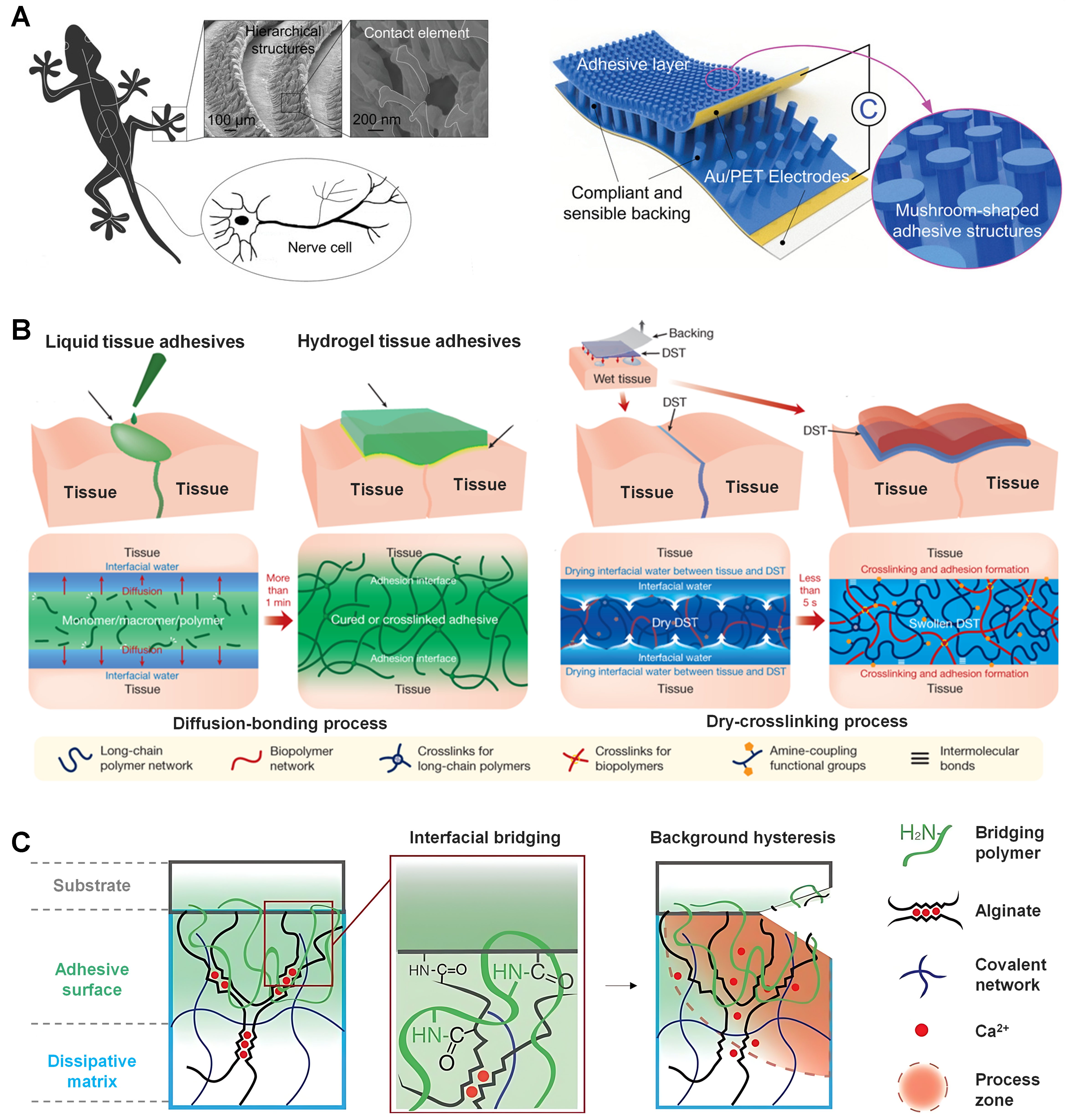
Figure 7. Bioinspired strategies and interfacial mechanisms for hydrogel-based tissue adhesion. (A) Bioinspiration from natural adhesive systems, where hierarchical surface structures such as gecko-inspired architectures provide design principles for contact-mediated adhesion across multiple length scales[183]. Copyright 2022, Wiley-VCH; (B) Bioinspired adhesive microstructures integrated into engineered devices, where mushroom-shaped microstructures combined with compliant substrates and conductive electrodes enable conformal contact and enhanced interfacial adhesion[186]. Copyright 2019, Springer Nature; (C) Mechanisms of hydrogel-based tissue adhesion, including polymer network formation, interfacial bridging, and molecular interactions such as covalent bonding, ionic coordination, and polymer entanglement that contribute to strong adhesion and energy dissipation at the tissue–material interface[187]. Copyright 2017, The American Association for the Advancement of Science. PET: Polyethylene terephthalate; DST: double-sided tape.
Chemical inspirations provide a complementary molecular route[190]. Networks rich in hydrogen bonds and aromatic groups interact with interfacial proteins and mucin glycoproteins, resembling transient biological bonding[191,192]. Polyphenols illustrate how metal coordination and dynamic covalent bonds can reinforce adhesion without sacrificing tolerance, allowing application-oriented balance between residence and safe removal[193]. Such adhesion must be defined by therapeutic outcomes rather than maximal strength at all costs.
Stability in extreme environments
Many conventional hydrogels rely on ester- or amide-based cross-linking pathways that can be vulnerable to hydrolysis in strongly acidic or high-salt media. Cleavage of cross-linking nodes within the backbone leads to decline in modulus, excessive swelling, and eventual structural failure. For example, Sun et al. showed that ionically crosslinked hydrogels rapidly lost mechanical integrity and adhesion under high ionic strength conditions due to charge screening and network dissociation[62]. Protonation and ionic screening at tissue interfaces further weaken electrostatic attraction and hydrogen-bonding, giving rise to short-lived adhesion and delamination[194]. These phenomena are particularly problematic in the gastrointestinal tract, sweat-rich chronic wounds, and the ocular surface dominated by tear fluids[195]. Addressing this challenge, Yang et al. developed a mucus-inspired hydrogel with protonation-driven adhesion that remained structurally stable and strongly adhesive under highly acidic conditions (pH ≈ 2), enabling effective esophageal tissue repair in vivo [Figure 8A][196]. Therefore, reliable performance requires dual or dynamically reinforced networks incorporating chemically inert motifs, so that resistance to chemical attack and maintenance of interfacial coupling can be achieved simultaneously.
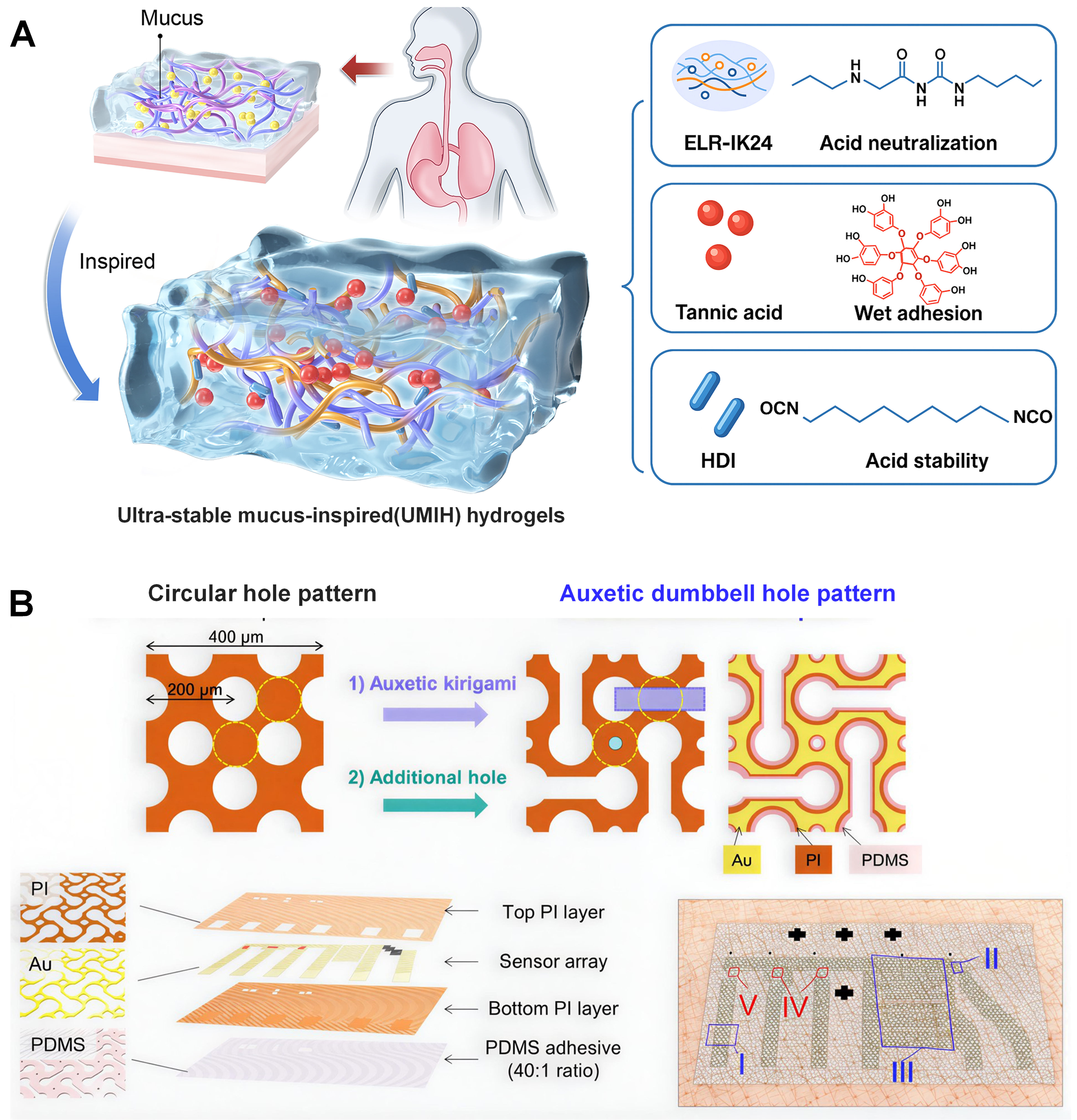
Figure 8. Bioinspired hydrogel design and electronic architectures for biointegrated systems. (A) Design of a mucus-inspired ultra-stable hydrogel integrating ELR-IK24 proteins, tannic acid, and HDI to achieve acid buffering, wet adhesion, and enhanced structural stability[196]. Copyright 2025, Elsevier; (B) Auxetic kirigami-based electronic architecture, where dumbbell-shaped kirigami patterns enable stretchable electronic interfaces integrating Au electrodes and PI layers on PDMS substrates for conformal biointegration and stable electrical performance[198]. Copyright 2021, The American Association for the Advancement of Science. HDI: Hexamethylene diisocyanate; PI: polyimide; PDMS: polydimethylsiloxane; ELR: elastin-like recombinant.
Encapsulation and evaporation suppression
Interface hydration must be preserved during exposure to air, as dehydration alters ionic mobility, interfacial impedance, and consequently sensing accuracy[197]. Along this line, Yeon et al. demonstrated that sweat pore-inspired perforated e-skins effectively mitigate sweat accumulation at the skin-device interface, thereby preserving conformal contact and long-term sensing stability during daily activities [Figure 8B][198]. Two complementary strategies are commonly used. Breathable yet waterproof polymeric membranes serve as physical barriers to evaporation while permitting gas and metabolite diffusion[199]. Hydrophobic or mucus-mimicking surface coatings provide additional regulation of interfacial water activity, enabling consistent permeability and signal stability under long-term wearing[200]. The combination of encapsulation engineering and biomimetic skins has emerged as a practical design principle for translating soft hydrogels into continuous monitoring platforms.
Multifunctional integration and system compatibility
Bio-interfaces are evolving from single adhesive layers to hubs for electrical and therapeutic exchange[201]. Conductive zones must support reliable percolation for sensors, while elastic networks demand low filler loading to preserve flexibility. Integration introduces challenges including sensing crosstalk and mismatch with flexible electronics or clinical workflows[202]. Therefore, molecular composition, microstructure, and encapsulation should be co-optimized to ensure predictable transport and stable system compatibility.
Progress toward clinical translation will rely on three dimensions. Programmable stimuli-responsive interfaces with reversible adhesion will address dynamic wet boundaries. AI-assisted prediction of interfacial lifetime could reduce empirical trial in material selection. Standardized testing protocols and long-term durability assessments will enable cross-laboratory comparison and regulatory evaluation. Convergence of these routes will determine how bioinspired soft interfaces mature into reliable wearable and implantable technologies.
EMERGING TRENDS AND CROSS-CUTTING APPLICATION SCENARIOS
Stimuli-responsive functionalization of soft hydrogels
Physical stimulus response
By incorporating photothermal nanomaterials (such as Mxene, polydopamine, and gold nanorods), hydrogels can efficiently convert light energy of specific wavelengths (primarily near-infrared) into heat[203], thereby inducing localized thermally triggered volume phase transitions, which in turn generate programmed bending, curling, or grasping motions[204,205]. Inspired by the phototropic behavior of
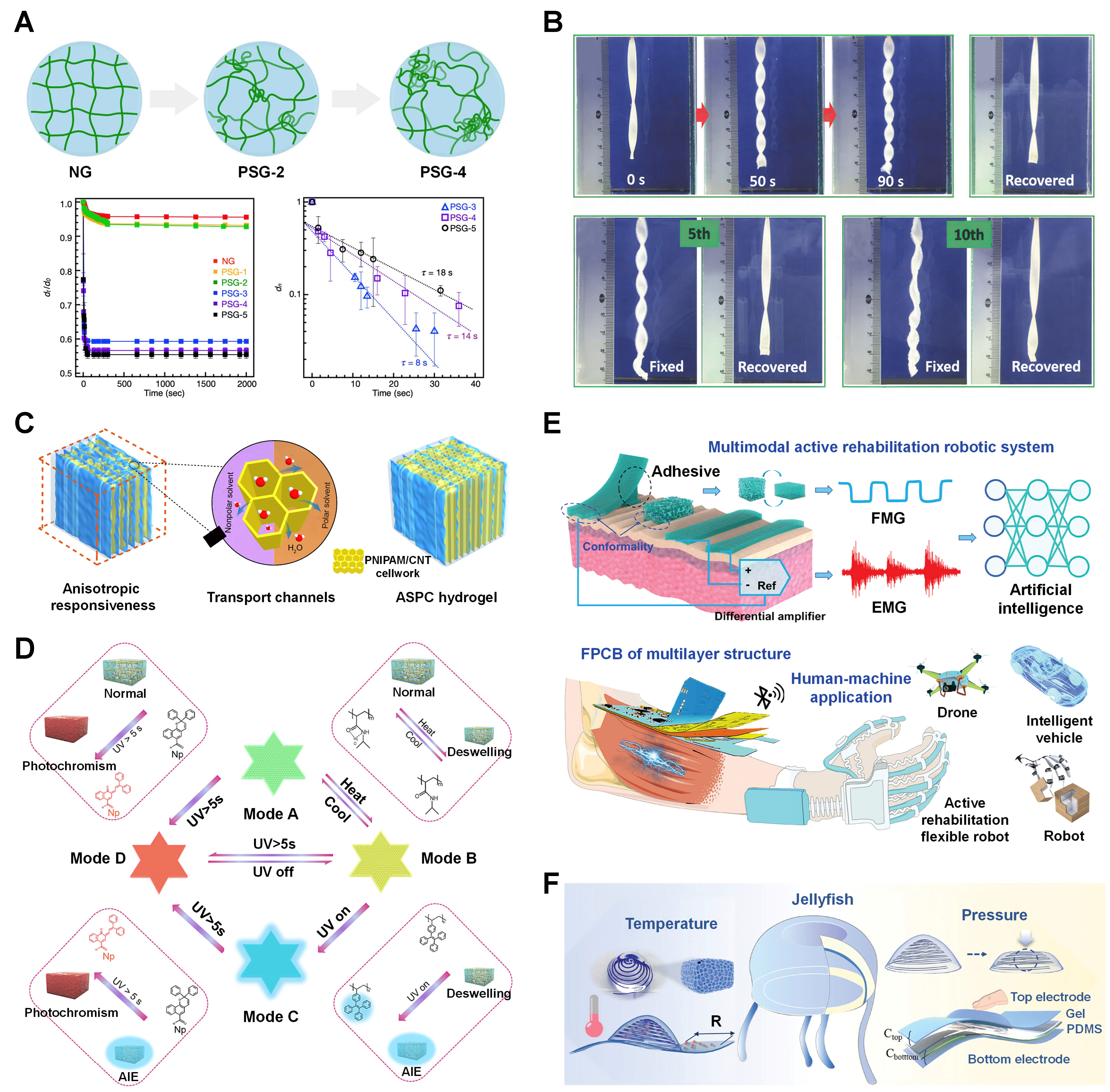
Figure 9. Stimuli-responsive mechanisms, responsivity, and multimodal sensing integration of soft hydrogels. (A) Network structures with phase separation and shrinking kinetics of normal hydrogel (NG) and phase-separated hydrogel (PSG)-1 to PSG-5 after a temperature jump from 20 to 45 °C, showing dn vs. time with relaxation times from the slopes[209]. Copyright 2021, MDPI; (B) Reversible twisting and recovery of M1S0.5A0.5 hydrogel in 0.1 M HCl and NaOH solutions, demonstrating consistent shape-memory behavior over at least 10 cycles[218]. Copyright 2018, Wiley-VCH; (C) Schematic of anisotropic ASPC hydrogel deformation in different solvents, facilitated by rapid water transport through low-tortuosity porous channels[219]. Copyright 2024, Springer Nature; (D) Multistage responsive behavior of CdS photonic crystal organohydrogel[228]. Copyright 2024, Elsevier; (E) The wearable human–machine interaction interface design using hydrogel-based EMG and pressure sensors for an AI-assisted active rehabilitation robotic system[259]. Copyright 2024, Wiley-VCH; (F) A jellyfish-inspired biomimetic hydrogel sensor device for temperature and pressure sensing[278]. Copyright 2025, Wiley-VCH. ASPC: Ag/Sa (sodium alginate)/PNIPAM [poly(N-isopropylacrylamide)]/CNT (carbon nanotube); EMG: electromyogram; PNIPAM: poly(N-isopropylacrylamide); CNT: carbon nanotube; UV: ultraviolet; AIE: aggregation-induced emission; FMG: force myography; FPCB: flexible printed circuit board; PDMS: polydimethylsiloxane.
Chemical stimulus response
Chemical responsiveness enables hydrogels to deeply interact with biological microenvironments[214,215]. Networks containing weak acidic/basic groups (e.g., carboxyl groups in polyacrylic acid (PAA) or amino groups in chitosan) exhibit significant swelling changes in response to environmental pH[216,217]. For example, Zhang et al. developed a polyampholyte hydrogel capable of spontaneous shape-memory cycles and macroscopic motion in response to alternating acidic/alkaline solutions, demonstrating its potential as a soft actuator [Figure 9B][33,218]. Recently, Yao et al. developed a solvent-adaptive hydrogel with a layered, constrained honeycomb structure [Figure 9C][219].
Stimulus-responsive mechanisms
Volume change represents the most fundamental stimulus-responsive behaviour of hydrogels, primarily driven by abrupt variations in osmotic pressure within the network or significant alterations in polymer-solvent interactions[220]. To transform uniform volumetric changes into directional, programmable deformations (such as bending, twisting, or curling), asymmetry or gradient structures must be introduced within the hydrogel to generate internal stress differences under stimulation[221,222]. For example, Wu et al. researched a novel soft hydrogel system where in-situ proton transfer enables programmable shape-morphing under light stimulation, independent of external water exchange[223]. Patterned printing of hydrogels with distinct responsiveness enables programmed two-dimensional to three-dimensional deformations[224]. Chen et al. developed a multiprogrammable anisotropic soft hydrogel by embedding magnetically oriented ferromagnetic nanoplates, which exhibits anisotropic volume response with distinct swelling ratios parallel and perpendicular to the alignment direction. This deliberate structural ordering enables the programmable integration of optical, mechanical, and magnetic properties for advanced soft actuators[225]. A typical example involves aligning magnetic nanoparticles along a single direction and fixing them within the network, so that the hydrogel bends predominantly in the vertical direction under magnetic or photothermal actuation[225]. Such preprogrammed anisotropy provides a powerful tool for achieving complex and predictable deformations.
Building on the concept of volume-dependent transformations, hydrogels can also demonstrate color responses through structural coloration mechanisms. The color response of hydrogels (structural coloration) does not originate from chemical dyes but rather from interference, diffraction, or scattering of light by their internal periodic nanostructures[226,227]. This coloration dynamically adjusts with changes in nanoscale spacing. For example, Chen et al. developed a photochromic organohydrogel that exhibits distinct color changes under UV light due to the reversible molecular conformational switching of naphthalene (Np) groups, enabling dynamic optical properties [Figure 9D][228]. When stimuli induce uniform swelling or shrinkage of the hydrogel[229], the lattice constant (interparticle spacing) changes accordingly, resulting in shifts in the wavelength of Bragg-diffracted light. Macroscopically, this manifests as continuous and reversible color changes. The quantitative relationship between color and stimulus intensity makes such hydrogels ideal candidates for visual sensing applications[229].
Expanding beyond optical responses, hydrogels can also exhibit photoelectric effects for energy conversion and sensing. Photoelectric response refers to the ability of materials to generate or modulate electrical signals (current, voltage, or resistance) under illumination[230]. Achieving efficient photoelectric response in hydrogels requires constructing a complete energy conversion pathway, from light harvesting to charge generation and subsequent charge transport[231]. Wu et al. developed a self-powered photoelectric sensor based on soft hydrogel diodes doped with photoacid, where the photoelectric response mechanism involves light-induced proton release from the photoacid and subsequent ion migration driven by the built-in electric field of the hydrogel PN junction[232]. The integrating photoelectric units into hydrogel networks could enable self-powered, soft bio-interfaces for wearable sensors or devices.
Flexible electronics integration and e-skin
Flexible e-skin platforms have been widely adopted in wearable and implantable biomedical devices for continuous health monitoring and therapeutic interfacing. In wearable applications, conformal e-skins can laminate onto the epidermis to monitor electrophysiological signals [electrocardiogram (ECG), electromyogram (EMG)][233], temperature[234], strain[235], and biochemical markers[236], supporting applications such as smart rings[237], epidermal patches[238], and soft rehabilitation monitors[239]. Their intimate skin contact reduces motion artifacts compared with rigid electronics, enabling higher signal-to-noise ratios during daily activities.
Implantable flexible electronics further push these advantages into in vivo environments, including neural probes[240], cardiac interfaces[241], and bioelectronic medicine systems. Ultrathin and mechanically compliant devices can follow tissue micromotions, reducing chronic inflammation and fibrotic encapsulation while maintaining stable electrical contact. In both wearable and implantable contexts, the integration of stretchable substrates, soft encapsulation layers, and biocompatible materials is critical for ensuring long-term safety, durability, and functional stability. A key design principle in e-skin integration is mechanical matching between electronic components and biological tissues. Flexible polymers[242], elastomers[243], and hydrogel-based substrates[244] are commonly used to achieve low modulus and high stretchability. Conductive elements are engineered using thin metal films[245], conductive polymers[246], liquid metals[247], or nanocomposites[248], often arranged in serpentine or mesh geometries to accommodate large strains without electrical failure. Multilayer architectures enable the decoupling of sensing, interconnection, and encapsulation functions, allowing for the integration of complex systems while preserving overall softness and conformability. Beyond materials, signal processing and system-level integration are central to the performance of e-skin systems. Soft bio-interfaces are continuously exposed to noise sources arising from motion, sweat, temperature fluctuations, and environmental electromagnetic interference. To address these challenges, e-skin platforms combine optimized electrode-tissue interfaces with low-noise front-end electronics, on-board amplification, and filtering to stabilize signal output[249]. At the interconnection level, bendable electrodes and stretchable interconnects maintain electrical continuity under repeated deformation, while flexible packaging strategies protect sensitive components from moisture and mechanical damage. At the system level, integrating sensing, data acquisition, power management, and wireless communication into compact, flexible architectures reduces parasitic noise and signal loss. The co-design of hardware and signal-processing algorithms enables adaptive noise suppression and artifact rejection, which is particularly important for long-term monitoring and closed-loop therapeutic systems[250]. Overall, flexible electronics integration provides the technological foundation for e-skin systems that are mechanically compliant, electrically stable, and functionally robust. Continued advances in soft materials, stretchable interconnects, and integrated signal processing will further expand the capabilities of e-skin, enabling multifunctional platforms for continuous health monitoring, neural interfacing, and next-generation wearable and implantable medical devices.
Sensor modalities and performance metrics
The central mission of flexible electronics and e-skin is to endow machines or prosthetics with sensory capabilities comparable to, or even surpassing, those of natural skin[251]. High-precision and reliable detection of diverse mechanical signals, including stretching, compression, bending, and their derivatives such as pressure and strain, is fundamental to achieving tactile perception, physiological monitoring, and motion feedback[252]. Owing to their soft mechanical properties matching biological tissues, tunable conductivity, and outstanding fracture toughness, soft hydrogels have emerged as ideal materials for constructing the next generation of durable flexible mechanical sensors[253,254]. Based on their working mechanisms, these sensors can be categorized into force/pressure sensors, strain sensors, and multifunctional integrated sensors, each designed to accurately detect tension, compression, bending, and pressure signals[255-258].
Force and pressure sensors are primarily employed to detect normal forces or pressures perpendicular to the device surface[255,256]. Wang et al. developed a multimodal hydrogel-based sensing system that integrates EMG and force myography (FMG) signals, leveraging a macroporous Foam-PAM hydrogel pressure sensor to detect subtle muscle forces for active rehabilitation in human-machine interfaces [Figure 9E][259]. In addition, Rahman et al. discovered that incorporating zeolitic imidazolate framework-8 (ZIF-8) nanoparticles into a poly(acrylamide)-co-hydroxyethyl acrylate (PAAm-co-HEA) hydrogel significantly enhances its force-responsive properties, enabling the fabrication of a highly stretchable triboelectric nanogenerator (TENG) that effectively converts biomechanical forces into electrical signals for self-powered sensing and energy harvesting[260]. Strain sensors are used to monitor stretching, compression, or bending of surfaces, with outputs typically correlated to strain (ε)[261]. Tensile strain detection is predominantly piezoresistive[262], when hydrogels are stretched, internal conductive pathways (such as ionic channels or networks of conductive nanofillers) are geometrically elongated and cross-sectional areas reduced, leading to increased resistance (positive strain effect). Soft hydrogels can withstand reversible tensile strains, thereby offering a broad detection range[254]. Their high toughness ensures that even under extreme stretching or localized damage, conductive networks remain continuous, allowing signals to recover. Compression sensing mechanisms overlap with those of normal pressure detection but emphasize volumetric deformation. Porous or foam-like hydrogels are particularly advantageous[263,264], upon compression, pore closure densifies conductive pathways, reducing resistance, while capacitance characteristics also change. Bending can be regarded as a composite state of tensile strain on one side and compressive strain on the other[261]. Sensors are typically attached to bendable surfaces, with resistance changes reflecting curvature. Critical factors include interfacial adhesion[265,266], ensuring hydrogels deform cooperatively with substrates without delamination.
Key performance indicators for sensors include sensitivity, detection range, response time, and stability[267-269]. Sensitivity quantifies the ability of sensors to convert mechanical stimuli (e.g., strain or pressure) into electrical signals (e.g., resistance or capacitance changes), determining resolution and amplification of small stimuli[267,269]. The high sensitivity of soft hydrogel sensors arises from the synergistic interplay between microstructural features and macroscopic mechanical properties[270]. Detection range is defined by the minimum (lower limit) and maximum (upper limit) stimuli that elicit effective responses. The broad elasticity and structural integrity of soft hydrogels provide a physical basis for extending detection ranges. For instance, a highly cracked hydrogel design that achieves multi-sensing with high sensitivity and a large detection range for soft machines by utilizing the opening and closing of pre-cut cracks to significantly alter current flow[271]. Response time characterizes the ability of sensors to track dynamic or transient mechanical stimuli[272], which is crucial for monitoring vibrations, speech, high-speed impacts, or physiological pulses. Benefiting from low hysteresis and porous architectures[263], soft hydrogels typically exhibit superior performance. Stability reflects the ability of sensors to maintain functionality under repeated use and environmental fluctuations, representing a core advantage of soft hydrogels[269]. Their energy dissipation mechanisms, fatigue resistance, and synthetic strategies conferring environmental robustness collectively determine sensor lifespan and reliability. Despite their excellent performance in mechanical sensing, soft hydrogel sensors still face multiple challenges in practical applications[83,273,274]. Conventional hydrogels undergo significant water-induced swelling in aqueous environments, leading to structural deformation, mechanical degradation, and signal instability. This limits their reliable use in complex conditions such as underwater sensing or high-humidity environments. In contrast to swelling, hydrogels are also prone to rapid moisture loss in ambient or dry conditions, leading to dehydration within hours. This compromises their mechanical integrity, ionic conductivity, and sensing functionality. Practical applications demand a synergy of features, such as mechanical robustness, high conductivity, self-adhesion, self-healing, biocompatibility, and antimicrobial properties, which are challenging to incorporate into a single hydrogel system.
One of the primary objectives of flexible sensors based on soft yet robust hydrogels is the construction of biomimetic, multifunctional e-skin[275,276]. An ideal e-skin system must not only transcend the detection of single mechanical signals but also achieve multimodal environmental perception[277]. Moreover, as an interface in long-term, intimate contact with biological systems, it must exhibit excellent wearing comfort, conformability, and biocompatibility. Owing to their tunable physicochemical properties, mechanical compatibility with tissues, and inherently hydrophilic interfaces, soft hydrogels represent an ideal material platform for this purpose. Recent advances can be reviewed from three perspectives, multimodal sensing integration, conformability, and biocompatibility.
E-skin must emulate the ability of natural skin to perceive multidimensional information, integrating synchronous or selective responses to stimuli such as temperature, humidity, chemical composition, and tactile signals. The design versatility of soft hydrogels provides unique advantages. Temperature sensing can exploit the temperature dependence of ionic conductivity in hydrogels (following the Arrhenius relationship), enabling a single conductive hydrogel to achieve self-sensing of temperature while monitoring mechanical signals. The working mechanism introduced by Ren et al. involves a temperature-sensing schematic where variations in temperature directly influence electrical resistance [Figure 9F][278]. This alteration in resistance is subsequently detected by an external circuit, facilitating temperature measurement. This well-defined linear dependence enables precise discrimination between different temperature levels. Humidity sensing relies on the hygroscopic nature of hydrogels, with electrical properties (e.g., ionic conductivity and dielectric constant) closely correlated to ambient humidity[279]. Ding et al. developed a groundbreaking self-powered, flexible chemosensor by engineering a smart hydrogel incorporated with zinc and lithium ions, enabling it to reversibly switch between water-rich and water-deficient states[280]. This single device achieves crosstalk-free detection of both oxygen and humidity with remarkable sensitivity (up to 4,170.5 %/% for O2 and 380.2 %/% RH for humidity) by leveraging a metal-air battery structure where the target molecules selectively act as limiting reactants or catalysts in the oxygen reduction reaction depending on the hydrogel’s state. Chemical sensing can be achieved by immobilizing specific recognition elements (e.g., glucose oxidase, aptamers, or molecularly imprinted polymers) within hydrogel networks. Inspired by human skin, Yun et al. developed a novel hydrogel-based sensing platform for two-dimensional imaging of external stimuli like chemicals[281]. This design enables high sensitivity 2D imaging through electrochemiluminescence, representing a significant advance toward biocompatible e-skins that leverage chemical reactions for multi-stimuli detection. Tactile sensing (pressure, strain, shear force) forms the foundation of multimodal perception, yet signal decoupling remains a central challenge. Tao et al. developed a self-powered tactile hydrogel sensor featuring a micro-pyramid-patterned double-network ionic organohydrogel, which detects subtle pressure changes through triboelectric signals without an external power supply[282]. This sensor exhibits remarkable sensitivity (45.97 mV·Pa-1), a fast response time (~20 ms), and operates reliably across a broad temperature range (-20 to 60 °C). Furthermore, its successful integration into a system that controls electronic devices and a robotic hand by mimicking finger gestures demonstrates significant potential for wearable electronics and human-machine interfaces. Comfort and skin-fitting characteristics are essential requirements for e-skin. Soft hydrogels, through precise tuning of mechanical properties and interfacial characteristics, provide key solutions to this requirement[283]. Chen et al. developed a wet-adaptive electronic skin (WADE-skin), highlighting that maintaining biological comfort at the device-skin interface in wet environments is crucial for the long-term, imperceptive wearability of epidermal electronics[284]. Zhang et al. developed a porous thermoplastic polyurethane (TPU)/carbon black hydrogel sensor via water vapor-induced phase separation, which exhibited high water vapor transmission rates
Biocompatibility is a paramount requirement for hydrogel-based e-skin to ensure safe, long-term wearability and prevent skin irritation or inflammatory responses, which are common issues with impermeable materials. Ma et al. developed a highly permeable and superelastic liquid-metal fibre mat (LMFM) that demonstrated excellent biocompatibility, as confirmed by in vitro cell viability tests showing over 95% cell survival and in vivo tests on rabbit and human skin showing no significant irritation, making it ideal for monolithic, multi-layered e-skin devices[287]. Additionally, Yan et al. developed conductive cellulose-based bio-nanosheet hydrogels by using polydopamine-reduced graphene oxide (GO) as a template for cellulose assembly. This approach resulted in a hydrogel that exhibits remarkable stability under physiological conditions while maintaining excellent electrical functionality and the ability to support cell growth, showcasing its high potential for creating long-term, bio-integrated electronic devices[288].
Soft robotics and actuators
Soft robotics has emerged as a transformative paradigm in robotics, offering mechanical compliance, adaptability, and safety, with relevance to minimally invasive surgery, wearable assistive devices, human-robot interaction, and unstructured environment exploration[289-291]. In contrast to rigid robots built from metals and hard polymers, soft robots are constructed from compliant materials capable of large deformation[292]. Their functionality relies on soft actuators that convert energy into controlled motion. Actuator performance depends on actuation mechanisms that govern responsiveness and operational constraints, and on structural design that determines motion complexity and controllability. This section reviews advances in soft actuators with emphasis on actuation principles and structural strategies enabling complex behaviors.
Actuation mechanisms
Soft actuators can be categorized based on stimulus type, including electrostatic, electroosmotic, thermal, photoinduced, moisture-responsive, and ion-driven systems.
Electrostatic actuation, exemplified by dielectric elastomer actuators (DEAs), relies on Maxwell stress generated by electric fields across dielectric layers between compliant electrodes[293]. Voltage application compresses the thickness and expands the in-plane area, enabling large strains[294]. These actuators offer high energy density, fast response, and silent operation, making them ideal for artificial muscles and dynamic morphing structures [Figure 10A][295], but often require high driving voltages, raising challenges for portability and safety, and may suffer from electrical breakdown and instability.
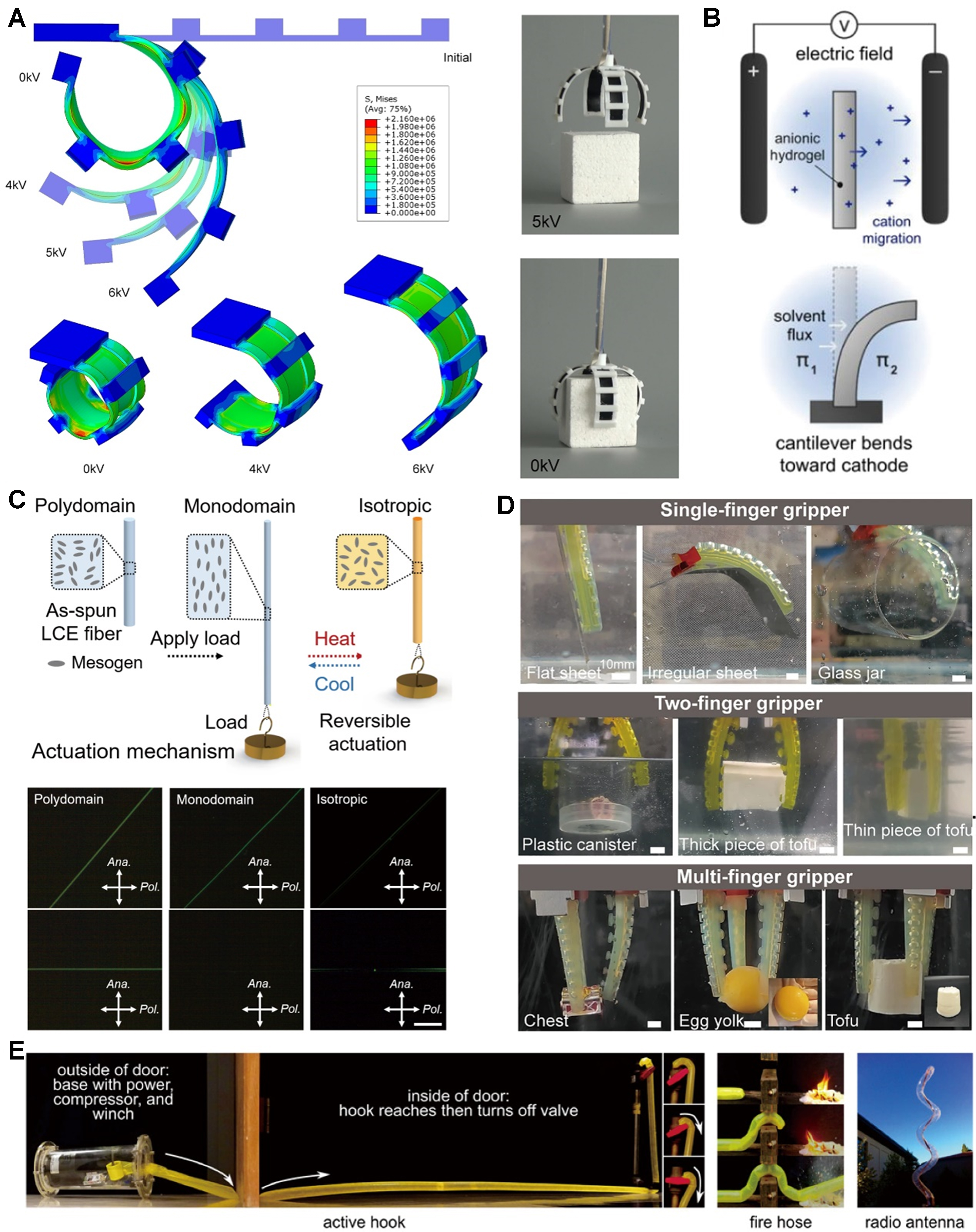
Figure 10. Actuation mechanism and motion complexity and structural design. (A) DEA soft gripper grips a cube[295]. Copyright 2019, Elsevier; (B) Illustration of electro-osmotic actuation mechanism[296]. Copyright 2022, American Chemical Society; (C) Top: Actuation mechanism of LCE fiber; bottom: POM images of polydomain, monodomain, and isotropic states of LCE microfibers observed at two different angles with respect to the analyzer[300]. Copyright 2021, The American Association for the Advancement of Science; (D) Designed and manufactured single-finger, two-finger, and multi-finger hydrogel soft grippers for handling objects of different shapes (spheres, cylinders, and cubes), surfaces (flat, curved, and folded) and stiffness attributes (stainless steel, glass, plastic, and ultra-soft foods like tofu and egg yolks)[314]. Copyright 2026, Springer Nature; (E) The soft robot demonstrates the ability to lengthen into useful 3D structures[315]. Copyright 2017, The American Association for the Advancement of Science. DEA: Dielectric elastomer actuator; LCE: liquid crystal elastomer; POM: polarized optical microscopic.
Electroosmotic actuation exploits the movement of ions and solvent in porous or hydrogel matrices under electric fields [Figure 10B][296]. It operates at low voltage and is suitable for micro-robot and microfluidic applications but can be limited by diffusion-constrained kinetics and modest force output. Thermally induced actuation relies on thermal expansion mismatch or phase transitions in materials such as shape memory polymers[297], liquid crystal elastomers[298], and bimorph composites[299]. Liquid crystal elastomers can contract anisotropically along mesogen alignment above phase transition temperatures, resembling muscle behavior [Figure 10C][300]. Thermal approaches provide large stroke and force but can be slow and energy-intensive. Photothermal agents such as CNTs, graphene, and gold nanoparticles can enable remote and spatially controlled heating under light[301]. Photoinduced actuation enables wireless control by converting light into mechanical work. Direct systems use molecular photoswitches such as azobenzene through trans cis isomerization[302], while indirect systems combine light absorbers with thermoresponsive matrices to achieve noncontact actuation[303]. These strategies are attractive for tether-free biomedical operation. Moisture-responsive actuation relies on differential swelling of hygroscopic materials such as cellulose under humidity gradients[304]. Biomimetic designs inspired by seed dispersal mechanisms can generate coiling, twisting, crawling, and other motions[305,306]. Such actuators can operate autonomously but may face slow response, limited reversibility in dry conditions, and durability concerns. Ion-driven actuation includes ionic polymer metal composites and conductive polymer actuators[307]. In ionic polymer metal composites, cation migration under low voltage bias causes hydration gradients and bending. These systems offer flexibility and bidirectional control[308] but may be limited by the solvent in electrolyte evaporation, electrode delamination, and finite lifetime.
Motion complexity and structural design
Structural architecture strongly influences motion richness and controllability. Advances in fabrication, including multi-material three-dimensional printing, soft lithography, and kerygma and origami-inspired patterning, have enabled sophisticated geometries. Basic kinematic modes include bending, contraction, and elongation, and twisting and helical motion. Bending can be achieved through asymmetric material distribution or gradient crosslinking and is widely used in grippers and locomotive units[309]. Contractile actuators based on pneumatic networks, DEAs, or liquid crystal elastomers generate linear motion for propulsion[294,298,309]. Twisting and helical motion can be produced through chiral winding or anisotropic alignment, enabling rotary propulsion in swimming microrobots[310,311]. Programmable motion can be achieved by patterning actuation domains for sequential or selective activation. A dual stimuli responsive hydrogel combining light and humidity sensitivity demonstrated autonomous sequence execution through environmental feedback[312]. Electrode arrays integrated into DEA membranes can generate traveling wave deformation[313]. Biomimetic structural designs include octopus inspired grippers with tapered chambers for adaptive grasping [Figure 10D][314] and vine tendril inspired elongating robots using localized swelling for tip extension [Figure 10E][315]. Layered architectures integrating sensing layers, actuator layers, and reinforcement elements enable closed loop systems with proprioception and adaptive response[316]. Embedded sensors based on stretchable conductors such as liquid metals and conductive hydrogels can monitor strain and pressure for feedback control[317,318]. A soft gripper equipped with resistive strain sensors, for example, can adjust gripping force based on object compliance, preventing damage[319].
Hydrogel actuators in varied environments
One particularly promising application of hydrogel actuators is in relatively complex multi-arm robots or grippers that can operate in a variety of environments[320-322]. These hydrogel actuators can grasp objects when exposed to certain stimuli and release them. Such responsive actuators are now extensively used as intelligent components in robotic systems, flexible sensing devices, and mobile medical or surgical biopsy instruments, largely due to their ability to be programmed into specific shapes. Although this line of application is still emerging, soft grippers that can grip and release objects are among the most promising developments. For example, to expand the range of robotic hand applications, by incorporating GO and polypyrrole (PPy), Dong et al. developed a programmable, patterned GO/PPy dual-responsive hydrogel actuator that responded efficiently to both temperature and infrared irradiation[323]. Remarkably, the gripper can lift objects 38 times its own weight, mimicking the way an eagle catches its prey. This research offers an effective approach for developing hydrogel actuators and paves the way for potentials in intelligent actuator technology.
A similar humidity-responsive hydrogel actuator was developed by Yao et al., who employed an Eu3+ ionic polyethyleneimine–acrylic acid copolymer (PEI-co-PAAC) in combination with poly(ethylene glycol) diacrylate (PEGDA)[324]. This actuator exhibited sensitivity to humidity and was capable of object detection and identification under ultraviolet light. Leveraging these properties, they designed a hexagonal hydrogel inspired by jellyfish camouflage, enabling the gripping and release of objects underwater. Zhao et al. reported another 4D printing innovation: an asymmetric bilayer-structured hydrogel actuator[325]. When gradually immersed in heated water, the actuator would catch and securely wrap a rubber block attached to a rope as immersion time increased. The wrapped object could then be moved by pulling the rope. Additionally, the actuator’s bending and releasing mechanism was adapted into bionic plant flower and hydrogel manipulator designs, making it easy to open/close or grasp/release target objects in response to thermal stimulation. Building on these strategies, researchers have recently developed even more advanced hydrogel actuators. These new designs function as intelligent fixtures, offering solutions that overcome the limitations of traditional fixtures, especially in challenging environments.
Thermoresponsive hydrogels are highly promising for use in soft actuators because they can reversibly change shape in response to temperature fluctuations[326,327]. Recent advance includes the development of tough, rapidly responding thermoresponsive DN hydrogels engineered for soft actuators by Zhang et al. The resulting hydrogels exhibit excellent mechanical performance, with an ultimate compressive stress of approximately 8 MPa[328]. They also display rapid actuation, reaching about 30% linear contraction and 28% radial contraction within 2 min. Furthermore, tubular soft actuators can serve as fluidic temperature sensors, switching the direction of fluid flow due to temperature variations.
Intelligent control and feedback system
The integration of intelligent control and feedback systems marks a major advance forward in the development of hydrogel-based soft robots. By embedding sensors and control units directly into the soft robotic structure, it becomes possible to continuously monitor key parameters such as fatigue, deformation, and actuation response in real time[329,330]. This closed-loop feedback allows for adaptive adjustments to actuator performance, ensuring reliable operation even in complex and changing environments. These smart systems not only improve the durability and functional reliability of hydrogel actuators but also broaden their potential for advanced applications in robotics, flexible sensing, and minimally invasive medical devices. Ultimately, combining responsive hydrogel materials with advanced control strategies sets the stage for the next generation of autonomous, adaptive, and multifunctional soft robotic systems[331,332]. For example, Huang et al. developed a foldable inductive sensor that could be integrated with an origami-inspired actuator[333]. With an increasing number of folds, the coil inductance decreased, leading to stable, sensitive, and highly repeatable performance, providing timely and reliable proprioceptive feedback for the gripper[333]. The final smart jellyfish design featured both buoyancy control and grasping capabilities under the water. In another example, Jin et al. developed a gas-driven flexible caterpillar robot equipped with advanced sensing capabilities[334]. The tactile sensors achieve a detection limit as low as 0.05 kPa along with an ultrafast response time of just 0.03 s. Meanwhile, the resistive strain sensors provide a sensitivity of 2.94 and can stretch up to 180%, allowing them to fully accommodate the robot’s body bending.
Quadrupedal soft robots also demonstrate unique advantages, such as rapid crawling speeds and versatile movement patterns, making them well-suited for tasks like navigation, obstacle avoidance, and exploration. For example, Xu et al. introduced a multifunctional, controlled-buckling sensor for monitoring the locomotion of a quadruped soft robot[335]. The robot’s body was constructed to be independently actuated along the grid in two directions, enabling movement within the first quadrant of a two-dimensional surface. In another work, Tang et al. advanced the field further by using 3D printing to create magnetic hydrogel soft structures capable of complex shape changes[336]. By incorporating a hard-magnetic filler into the hydrogel matrix and employing nano-colloids to tune the rheology of the precursor, they were able to directly print intricate magnetic hydrogel shapes. These printed components were assembled into a 3D-printed magnetic hydrogel lotus, which is fully bloomed without a magnetic field. Under a uniform magnetic field, the lotus spreads flat; an upward field causes it to close completely, while a downward field induces opening.
From multimodal biosensing to AI-enabled health interfaces
As hydrogel bioelectronics evolve from single-mode sensors to integrated health platforms, the key challenge is no longer only signal acquisition, but also the extraction of clinically meaningful information from complex, multimodal datasets. In this context, the convergence of hydrogel-based biosensing with wireless communication, microfluidic sampling, and AI-assisted data analysis is enabling a new generation of intelligent health interfaces capable of continuous monitoring, pattern recognition, and closed-loop intervention.
Wearable soft hydrogel biosensors
Soft hydrogels are pivotal for wearable biosensors, offering skin-like compliance, high signal fidelity, and multimodal sensing capabilities by seamlessly integrating with biological tissues. In recent years, research on wearable soft hydrogel biosensors has shifted from merely enhancing mechanical performance to achieving more stable, precise, and intelligent health signal acquisition. Pan et al. developed a MXene-functionalized PEDOT:PSS conductive hydrogel for non-invasive sweat glucose monitoring, demonstrating high conductivity and stability enabled by the material’s tailored porous network [Figure 11A][337]. Similarly, Zhao et al. engineered a dual-network hydrogel architecture with allylated hydroxyethyl cellulose with poly(3,4-ethylenedioxythiophene):poly(sodium-p-styrenesulfonate) and PAM, achieving remarkable pressure sensitivity and fast response for human motion tracking without external power[338]. Additionally, Wang et al. designed a rapid-polymerization hydrogel exhibiting autonomous self-healing and stable adhesion, allowing reliable operation under mechanical deformation and varying environmental conditions[339]. Furthermore, Shan et al. created an injectable hydrogel with integrated bacteria theranostic and motion monitoring functions, enabling real-time detection of joint movements and antibacterial protection for wound healing applications [Figure 11B][340]. These advances highlight how soft hydrogels, through rational multiscale design, are evolving from passive conductors to intelligent, multifunctional interfaces for continuous health monitoring. Despite these advances, soft hydrogel-based wearable biosensors are hindered by inherent material vulnerabilities, including environmental instability and signal interference from calibration drift and biofouling[341,342]. These limitations collectively challenge their long-term reliability and commercial viability for continuous health monitoring.
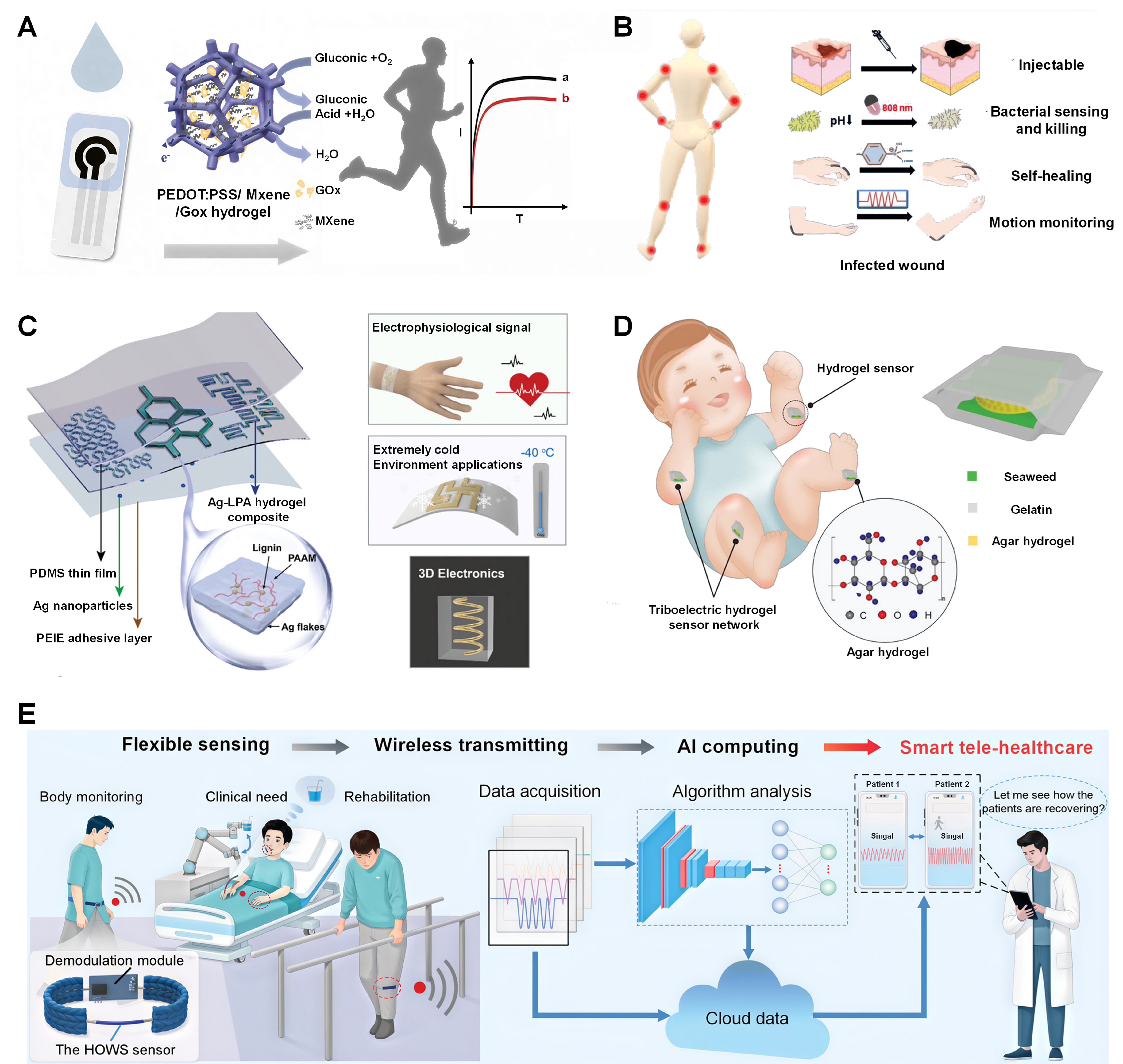
Figure 11. Representative design and applications of wearable hydrogel sensors for health monitoring. (A) demonstration of wearable sensor fabrication and sweat glucose detection[337]. Copyright 2024, Elsevier; (B) Structure, applications, and multi-functions of aminophenylboronic acid grafted SA (Alg-PBA)/PVA/GOH hydrogels[340]. Copyright 2024, Wiley-VCH; (C) The programmable microfluidic-assisted hydrogel patches[345]. Copyright 2024, Wiley-VCH; (D) A soft edible triboelectric hydrogel sensor for infant motion monitoring[346]. Copyright 2022, Wiley-VCH; (E) HOWS sensor featuring hydrogel optical waveguide with integrated wireless sensing and AI computing[347]. Copyright 2025, Wiley-VCH. SA: Sodium alginate; PBA: 3-aminophenylboronic acid; PVA: poly(vinyl alcohol); GOH: hydroxylated graphene; HOWS: hydrogel-based optical waveguide stretchable; PEDOT:PSS: poly(3,4-ethylenedioxythiophene):poly(styrenesulfonate); GOx: glucose oxidase; PDMS: polydimethylsiloxane; PEIE: polyethyleneimine; LPA: lignin-polyacrylamide; PAAM: polyacrylamide acrylamide.
Multimodal biosensing for comprehensive health data acquisition
Multimodal biosensing integrates diverse physiological signals to provide comprehensive health monitoring, enabling real-time assessment of both biochemical and biophysical parameters for personalized healthcare. Soft hydrogels, with their intrinsic flexibility, biocompatibility, and tunable conductivity, serve as ideal platforms for developing wearable sensors that can conform to biological tissues and facilitate simultaneous data acquisition from multiple sources. For instance, Imani et al. developed a hybrid chemical-electrophysiological sensing system that concurrently monitors sweat lactate and electrocardiogram (ECG) signals, utilizing hydrogel-based interfaces to ensure skin compatibility and stable signal transmission during physical activities[343]. Lin et al. developed a non-invasive sweat glucose sensor that concurrently monitors ECG signals, demonstrating the integration of chemical and electrophysiological sensing in a single wearable platform. This hybrid system allows for the correlation of metabolic activity with cardiac function, providing a more holistic health assessment[344]. Furthermore, Liao et al. engineered a programmable microfluidic-assisted hydrogel patch with a silver-lignin-PAM composite for monitoring physiological signals like heart rate and pulse. This approach highlights the use of soft hydrogels in creating structured, multifunctional sensing interfaces through advanced fabrication techniques [Figure 11C][345]. However, the broader deployment of soft hydrogel-based multimodal sensing systems is currently challenged by achieving environmental stability and integrated multimodal sensing for reliable clinical translation.
Data processing and AI-driven health insights
Soft hydrogels serve as an ideal platform for AI-driven health monitoring by providing high-quality, multimodal physiological data through their biocompatible and conformable interfaces. The integration of deep learning algorithms with these sensors addresses challenges in processing complex, noisy data, enabling intelligent diagnosis and prediction. Guo et al. developed a deep learning-assisted triboelectric hydrogel sensor that achieved 100% recognition accuracy in monitoring infant movements by analyzing signals from various activities. This system enables real-time safety monitoring through wireless data transmission to a mobile device [Figure 11D][346]. Li et al. engineered a hydrogel-based optical waveguide stretchable sensor integrated with a convolutional neural network to differentiate between normal and abnormal gait patterns for remote patient monitoring. This smart tele-healthcare system facilitates multimodal human-machine interaction, serving as a communication tool for patients with speech impairments [Figure 11E][347]. However, a key limitation in current systems is the need for extensive, individualized data pre-training to overcome signal variability and achieve robust model generalization for reliable clinical insights.
OUTLOOK AND FUTURE OPPORTUNITIES
Recent advances in soft yet robust hydrogels have opened new horizons for bio-integrated devices. To move beyond laboratory prototypes toward clinical translation and commercial adoption, future research must not only address reliability and stability but also explore transformative opportunities at the interface of materials science, engineering, and data intelligence.
Toward predictive reliability: Future studies should integrate accelerated aging models with in-situ monitoring to quantitatively predict hydrogel device lifespan. Coupling fatigue mechanics with multi-scale imaging (e.g., synchrotron X-ray tomography, cryo-EM) will reveal crack initiation and conductive network degradation in real time. Such mechanistic insights can establish predictive design rules for long-term operation under physiological stress.
Toward adaptive biointerfaces: Next-generation hydrogel interfaces should actively respond to biological cues rather than passively endure them. Smart surfaces capable of modulating immune reactions, releasing therapeutic agents, or dynamically adjusting adhesion will transform device–tissue integration. Incorporating bioinspired architectures (e.g., hierarchical porosity, anisotropic modulus gradients) can minimize fibrous capsule formation and achieve “invisible” integration.
Toward multimodal intelligence: Hydrogel platforms are poised to evolve into multifunctional systems that combine sensing, stimulation, and therapeutic delivery. Embedding machine learning into signal processing will allow cross-validation of diverse physiological inputs, advancing toward closed-loop diagnostic and therapeutic frameworks. Beyond ECG or EMG, multimodal hydrogel arrays could integrate biochemical sensing of metabolites, enabling personalized monitoring at the molecular level.
Toward scalable and sustainable translation: Commercialization requires convergence of scalable manufacturing, regulatory alignment, and sustainability. Roll-to-roll and 3D printing can reduce costs, while biodegradable polymers and circular design strategies will minimize environmental impact. AI-driven inverse design of hydrogel formulations, informed by quantitative composition–structure–property databases, will accelerate material discovery. Green manufacturing processes and modular device architectures will further establish a closed-loop material cycle, ensuring both clinical relevance and ecological responsibility.
CONCLUSIONS
Strong and resilient soft hydrogels have become essential materials for developing flexible bio-integrated devices, thanks to their unique blend of mechanical strength, biocompatibility, and customizable physical properties. Their true value lies in the ability to design hydrogels at multiple scales, from molecular structures and network configurations to larger assemblies, which allows for simultaneous improvements in toughness, resistance to fatigue, and stability in various environments. These multi-scale mechanical design approaches are crucial for ensuring that hydrogels can reliably function and integrate with both electronic components and biological tissues over the long term. Advanced fabrication and scalable manufacturing techniques, such as high-resolution 3D printing, microfabrication, and advanced interface packaging, make it possible to precisely control hydrogel characteristics and seamlessly incorporate them into complex device systems. These breakthroughs have driven major advancements in areas like wearable electronics, e-skin, soft robotics, and multimodal biosensors, where hydrogels play a key role in connecting materials engineering with biomedical applications.
Looking ahead, the field is on the verge of major breakthroughs fueled by trends like AI-driven material design, the creation of smart and adaptive interfaces, and the push for sustainable, scalable commercialization. These directions not only promise to speed up the discovery of next-generation hydrogel materials with exceptional performance, but also tackle key challenges related to clinical application and environmental sustainability. By combining data-driven design with cutting-edge fabrication and thorough in vivo testing, we aim to develop robust hydrogel platforms that can become clinically practical and environmentally friendly solutions for precision medicine and continuous health monitoring. In summary, the ongoing progress in strong and tough hydrogels, driven by multi-scale design and innovative manufacturing, will be crucial for enabling flexible, reliable, and intelligent bio-integrated devices that shape the future of healthcare.
Strong and resilient soft hydrogels have become key materials for flexible bio-integrated devices because they combine tissue-like softness, biocompatibility, and mechanically robust performance. Recent progress shows that multiscale design, from molecular networks to microstructures and device interfaces, is essential for improving toughness, fatigue resistance, and long-term stability under physiological conditions. In parallel, advances in fabrication and integration strategies have expanded the use of hydrogels in wearable electronics, e-skin, soft robotics, and biosensing systems.
Looking forward, further progress will depend on the integration of AI-assisted material design, adaptive bio-interfaces, scalable manufacturing, and rigorous in vivo validation. These efforts are expected to accelerate the development of hydrogel systems that are not only high-performing and reliable, but also clinically practical and environmentally sustainable. Overall, continued innovation in robust soft hydrogels will play a central role in enabling the next generation of flexible and intelligent bio-integrated devices for precision medicine and continuous health monitoring.
DECLARATIONS
Authors’ contributions
Conceptualization: Yang, K.; Liu, W.
Writing: Yang, K; Yang, X.; Zhu, W.; Liang, X.
Review and editing: Yang, K; Yang, X.; Zhu, W.; Lu, C.; Liang, X.
Supervision: Yang, X.; Zhu, W.; Liang, X.
Project assistance: Liu, W.; Yi, Z.
All authors contributed to the editing and approved the final version of the manuscript.
All authors have read and agreed to the published version.
Availability of data and materials
Not applicable.
AI and AI-assisted tools statement
During the preparation of this manuscript, OpenAI GPT-4.5 (accessed in December 2025) was used solely to assist with the generation and refinement of graphical elements for Figure 1 based on author-provided concepts and prompts. The tool did not influence the scientific content, experimental design, data collection, data analysis, interpretation, or conclusions of the work. All authors take full responsibility for the accuracy, integrity, originality, and final content of the graphical abstract and the manuscript.
Financial support and sponsorship
This work was supported by the National Natural Science Foundation (52403174); InnoHK initiative of the Innovation and Technology Commission of the Hong Kong roomSpecial Administrative Region Government; Natural Science Foundation of Guangdong Province (2025A0505010014, 2020A1515110288, 2025A1515011154); Natural Science Foundation of Hunan Province (2023JJ40655, 2025JJ40038); Shenzhen Science and Technology Program (RCBS20210609103713046, JCYJ20250604191212016); Agricultural Science and Technology Innovation Program and Outstanding Young Talents Funding of the Chinese Academy of Agricultural Sciences (CAAS-ASTIP-IBFC); Postdoctoral Research Start-up Funds of Dapeng New District and Shenzhen City.
Conflicts of interest
All authors declared that there are no conflicts of interest.
Ethical approval and consent to participate
Not applicable.
Consent for publication
Not applicable.
Copyright
© The Author(s) 2026.
REFERENCES
1. Cho, H.; Lee, I.; Jang, J.; et al. Real-time finger motion recognition using skin-conformable electronics. Nat. Electron. 2023, 6, 619-29.
2. Liu, H.; Xie, J.; Zhao, J.; et al. Construction of gradient SA-TiO2 hydrogel coated PVDF-g-IL fibre membranes with high hydrophilicity and self-cleaning for the efficient separation of oil-water emulsion and dye wastewater. J. Membr. Sci. 2024, 697, 122580.
3. Kim, K. Y.; Kang, J.; Song, S.; et al. An ultrathin organic–inorganic integrated device for optical biomarker monitoring. Nat. Electron. 2024, 7, 914-23.
4. Duan, Y.; Li, B.; Yang, K.; et al. Ultrahigh energy and power density in Ni-Zn aqueous battery via superoxide-activated three-electron transfer. Nanomicro. Lett. 2024, 17, 79.
5. Zhu, W.; Chow, L.; Ye, D.; et al. Advances in smart textiles for personal thermal management. Med-X 2025, 3, 50.
6. Shi, J.; Kim, S.; Li, P.; et al. Active biointegrated living electronics for managing inflammation. Science 2024, 384, 1023-30.
7. Li, X.; Xu, R.; Xie, C.; Ge, Z.; Gao, B.; Lim, C. T. Microscale architectures for intelligent soft robotics: from functional microneedles to biointegrated wearable systems. Nanomicro. Lett. 2026, 18, 179.
8. Yang, K.; Li, B.; Ma, Z.; et al. Ion-selective mobility differential amplifier: enhancing pressure-induced voltage response in hydrogels. Angew. Chem. Int. Ed. Engl. 2025, 64, e202415000.
9. Lu, Z.; Cui, J.; Liu, F.; et al. A 4D printed adhesive, thermo-contractile, and degradable hydrogel for diabetic wound healing. Adv. Healthc. Mater. 2024, 13, e2303499.
10. Mahato, K.; Saha, T.; Ding, S.; Sandhu, S. S.; Chang, A.; Wang, J. Hybrid multimodal wearable sensors for comprehensive health monitoring. Nat. Electron. 2024, 7, 735-50.
11. Sedighi, A.; Kou, T.; Huang, H.; Li, Y. Noninvasive on-skin biosensors for monitoring diabetes mellitus. Nanomicro. Lett. 2025, 18, 16.
12. Shi, J.; Li, P.; Kim, S.; Tian, B. Implantable bioelectronic devices for photoelectrochemical and electrochemical modulation of cells and tissues. Nat. Rev. Bioeng. 2025, 3, 485-504.
13. Oh, S.; Jekal, J.; Liu, J.; et al. Bioelectronic implantable devices for physiological signal recording and closed‐loop neuromodulation. Adv. Funct. Mater. 2024, 34, 2403562.
14. Yao, Y.; Huang, W.; Chen, J.; et al. Flexible and stretchable organic electrochemical transistors for physiological sensing devices. Adv. Mater. 2023, 35, e2209906.
15. Huang, L.; Tang, D.; Yang, Z. Flexible electronic materials and devices toward portable immunoassays. FlexMat 2024, 1, 59-78.
16. Wang, D.; Zeng, Z.; Ma, Z.; et al. Soft self-powered solid-state poly(ionic liquid) piezoionic elastomer sensor for extreme temperature operation. Chem. Eng. J. 2026, 530, 173003.
17. Zhang, Y.; Tan, Y.; Lao, J.; Gao, H.; Yu, J. Hydrogels for flexible electronics. ACS. Nano. 2023, 17, 9681-93.
18. Yang, G.; Qiu, Y.; Pang, B.; et al. A reusable hydrogel biosensor array with electrically responsive hydrogel interfaces for noninvasive locating of perforating arteries. Sci. Adv. 2025, 11, eadw6166.
19. Chong, J.; Sung, C.; Nam, K. S.; et al. Highly conductive tissue-like hydrogel interface through template-directed assembly. Nat. Commun. 2023, 14, 2206.
20. Gu, Y.; Luo, Y.; Guo, Q.; et al. Empowering human‐machine interfaces: self‐powered hydrogel sensors for flexible and intelligent systems. Adv. Funct. Mater. 2026, 36, e09085.
21. Lee, M. A.; Jin, X.; Muthupalani, S.; Bakh, N. A.; Gong, X.; Strano, M. S. In-vivo fluorescent nanosensor implants based on hydrogel-encapsulation: investigating the inflammation and the foreign-body response. J. Nanobiotechnology. 2023, 21, 133.
22. Zhan, Y.; Kim, S.; Song, Y.; et al. Wearable optical–electrical skin sensing platform for sweat gland dynamics monitoring. Nat. Sens. 2026, 1, 494-501.
23. Roy, A.; Zenker, S.; Jain, S.; et al. A highly stretchable, conductive, and transparent bioadhesive hydrogel as a flexible sensor for enhanced real-time human health monitoring. Adv. Mater. 2024, 36, e2404225.
24. Wang, Y.; Wang, Z.; Zhang, Y.; et al. A 2.7-μm-thick robust, permeable, and antifreezing hydrogel electrode for long-term ambulatory health monitoring. Sci. Adv. 2025, 11, eadt2286.
25. Shan, L.; Xue, Y.; Chen, X.; et al. Mechanically compliant and impedance matching hydrogel bioelectronics for low-voltage peripheral neuromodulation. Adv. Mater. 2026, 38, e11014.
26. Ding, J.; Chen, Z.; Liu, X.; et al. A mechanically adaptive hydrogel neural interface based on silk fibroin for high-efficiency neural activity recording. Mater. Horiz. 2022, 9, 2215-25.
27. Zhou, Y.; Jia, X.; Pang, D.; et al. An integrated Mg battery-powered iontophoresis patch for efficient and controllable transdermal drug delivery. Nat. Commun. 2023, 14, 297.
28. Mendez, K.; Whyte, W.; Freedman, B. R.; et al. Mechanoresponsive drug release from a flexible, tissue-adherent, hybrid hydrogel actuator. Adv. Mater. 2024, 36, e2303301.
29. Hu, M.; Ren, J.; Pan, Y.; et al. Scaled elastic hydrogel interfaces for brain electrophysiology. Adv. Funct. Mater. 2024, 34, 2407926.
30. Wang, J.; Wang, T.; Liu, H.; et al. Flexible electrodes for brain-computer interface system. Adv. Mater. 2023, 35, e2211012.
31. Alamdari, S. G.; Alibakhshi, A.; de la Guardia, M.; et al. Conductive and semiconductive nanocomposite-based hydrogels for cardiac tissue engineering. Adv. Healthc. Mater. 2022, 11, e2200526.
32. Ding, T.; Ren, C.; Meng, L.; et al. Nonswelling lubricative nanocolloidal hydrogel resistant to biodegradation. Nanomicro. Lett. 2025, 17, 327.
33. Lei, K.; Chen, M.; Guo, P.; et al. Environmentally adaptive polymer hydrogels: maintaining wet‐soft features in extreme conditions. Adv. Funct. Mater. 2023, 33, 2303511.
34. Li, X.; Gong, J. P. Design principles for strong and tough hydrogels. Nat. Rev. Mater. 2024, 9, 380-98.
35. Sun, X.; Mao, Y.; Yu, Z.; Yang, P.; Jiang, F. A biomimetic “salting out-alignment-locking” tactic to design strong and tough hydrogel. Adv. Mater. 2024, 36, e2400084.
36. Fang, C.; Gao, Z.; Chung, K. Y.; et al. Advanced design for electrochemically flexible iontronic tactile sensing. Adv. Compos. Hybrid. Mater. 2026, 9, 1829.
37. Petelinšek, N.; Mommer, S. Tough hydrogels for load-bearing applications. Adv. Sci. 2024, 11, e2307404.
38. Wang, S.; Lei, L.; Tian, Y.; et al. Strong, tough and anisotropic bioinspired hydrogels. Mater. Horiz. 2024, 11, 2131-42.
39. Chen, Y.; Li, J.; Lu, J.; Ding, M.; Chen, Y. Synthesis and properties of poly(vinyl alcohol) hydrogels with high strength and toughness. Polym. Test. 2022, 108, 107516.
40. Kim, J.; Zhang, G.; Shi, M.; Suo, Z. Fracture, fatigue, and friction of polymers in which entanglements greatly outnumber cross-links. Science 2021, 374, 212-6.
41. Pan, J.; Gao, L.; Sun, W.; Wang, S.; Shi, X. Length effects of short alkyl side chains on phase-separated structure and dynamics of hydrophobic association hydrogels. Macromolecules 2021, 54, 5962-73.
42. Qian, X.; Mu, N.; Zhao, X.; et al. Novel self-healing and recyclable fire-retardant polyvinyl alcohol/borax hydrogel coatings for the fire safety of rigid polyurethane foam. Soft. Matter. 2023, 19, 6097-107.
43. Zhou, X.; Li, T.; Wang, J.; et al. Shape morphing of anisotropy-encoded tough hydrogels enabled by asymmetrically-induced swelling and site-specific mechanical strengthening. J. Mater. Chem. B. 2018, 6, 4731-7.
44. Wang, S.; Chen, Y.; Sun, Y.; et al. Stretchable slide-ring supramolecular hydrogel for flexible electronic devices. Commun. Mater. 2022, 3, 225.
45. He, G.; Lei, H.; Sun, W.; et al. Strong and reversible covalent double network hydrogel based on force-coupled enzymatic reactions. Angew. Chem. Int. Ed. Engl. 2022, 61, e202201765.
46. Kedzierski, A.; Kheirabadi, S.; Jaberi, A.; et al. Engineering the hierarchical porosity of granular hydrogel scaffolds using porous microgels to improve cell recruitment and tissue integration. Adv. Funct. Mater. 2025, 35, 2417704.
47. Eckstein, K. N.; Hergert, J. E.; Uzcategui, A. C.; et al. Controlled mechanical property gradients within a digital light processing printed hydrogel-composite osteochondral scaffold. Ann. Biomed. Eng. 2024, 52, 2162-77.
48. Geng, L.; Liu, W.; Fan, B.; et al. Anisotropic double-network hydrogels integrated superior performance of strength, toughness and conductivity for flexible multi-functional sensors. Chem. Eng. J. 2023, 462, 142226.
49. Lv, Z.; Hu, T.; Bian, Y.; et al. A MgFe-LDH nanosheet-incorporated smart thermo-responsive hydrogel with controllable growth factor releasing capability for bone regeneration. Adv. Mater. 2023, 35, e2206545.
50. Fragiadakis, D.; Roland, C. M. Chain flexibility and the segmental dynamics of polymers. J. Phys. Chem. B. 2019, 123, 5930-4.
51. Fengler, C.; Keller, J.; Ratzsch, K. F.; Wilhelm, M. In situ RheoNMR correlation of polymer segmental mobility with mechanical properties during hydrogel synthesis. Adv. Sci. 2022, 9, e2104231.
52. Nian, G.; Kim, J.; Bao, X.; Suo, Z. Making highly elastic and tough hydrogels from doughs. Adv. Mater. 2022, 34, e2206577.
53. Xu, L.; Qiao, Y.; Qiu, D. Coordinatively stiffen and toughen hydrogels with adaptable crystal-domain cross-linking. Adv. Mater. 2023, 35, e2209913.
54. Mayumi, K.; Ito, K. Structure and dynamics of polyrotaxane and slide-ring materials. Polymer 2010, 51, 959-67.
55. Wang, X.; Liu, H.; Gao, M. Harnessing sacrificial bond kinetics for hydrogel self-strengthening. Innovation 2025, 6, 100988.
56. Wang, Z.; Xu, X.; Xu, Y.; Lin, W.; Peng, Z. A ternary heterogeneous hydrogel with strength elements for resilient, self-healing, and recyclable epidermal electronics. npj. Flex. Electron. 2022, 6, 175.
57. Okay, O. Self-healing hydrogels formed via hydrophobic interactions. In: Seiffert, S.; editor. Supramolecular polymer networks and gels. Cham: Springer International Publishing; 2015. pp. 101-42.
58. Hu, X.; Vatankhah-Varnoosfaderani, M.; Zhou, J.; Li, Q.; Sheiko, S. S. Weak hydrogen bonding enables hard, strong, tough, and elastic hydrogels. Adv. Mater. 2015, 27, 6899-905.
59. Wang, G.; Liu, Y.; Zu, B.; et al. Reversible adhesive hydrogel with enhanced sampling efficiency boosted by hydrogen bond and van der Waals force for visualized detection. Chem. Eng. J. 2023, 455, 140493.
60. Deng, S.; He, S.; Yan, G.; et al. π-π Conjugated bonds stacking/scattering for switchable lubrication in supramolecular hydrogel. Adv. Sci. 2025, 12, e2500447.
61. Gong, J. P.; Katsuyama, Y.; Kurokawa, T.; Osada, Y. Double‐network hydrogels with extremely high mechanical strength. Adv. Mater. 2003, 15, 1155-8.
62. Sun, J. Y.; Zhao, X.; Illeperuma, W. R.; et al. Highly stretchable and tough hydrogels. Nature 2012, 489, 133-6.
63. Zhang, W.; Hu, J.; Tang, J.; et al. Fracture toughness and fatigue threshold of tough hydrogels. ACS. Macro. Lett. 2019, 8, 17-23.
64. Okumura, Y.; Ito, K. The polyrotaxane gel: a topological gel by figure-of-eight cross-links. Adv. Mater. 2001, 13, 485-7.
65. Yasui, T.; Zheng, Y.; Nakajima, T.; Kamio, E.; Matsuyama, H.; Gong, J. P. Rate-independent self-healing double network hydrogels using a thixotropic sacrificial network. Macromolecules 2022, 55, 9547-57.
66. Riffe, M. B.; Davidson, M. D.; Seymour, G.; et al. Multi-material volumetric additive manufacturing of hydrogels using gelatin as a sacrificial network and 3D suspension bath. Adv. Mater. 2024, 36, e2309026.
67. Liang, X.; Qi, Y.; Pan, Z.; et al. Design and preparation of quasi-spherical salt particles as water-soluble porogens to fabricate hydrophobic porous scaffolds for tissue engineering and tissue regeneration. Mater. Chem. Front. 2018, 2, 1539-53.
68. Cao, P.; Chen, B.; Cao, Y.; Gao, H. Micropores can enhance the intrinsic fracture energy of hydrogels. Soft. Matter. 2025, 21, 2355-62.
69. Liang, X.; Chen, G.; Lin, S.; et al. Anisotropically fatigue-resistant hydrogels. Adv. Mater. 2021, 33, e2102011.
70. Liang, X.; Duan, P.; Gao, J.; et al. Bilayered PLGA/PLGA-HAp composite scaffold for osteochondral tissue engineering and tissue regeneration. ACS. Biomater. Sci. Eng. 2018, 4, 3506-21.
71. Wang, Y.; Qin, X.; Feng, Y.; et al. Dual-gradient silk-based hydrogel for spatially targeted delivery and osteochondral regeneration. Adv. Mater. 2025, 37, e2420394.
72. Gao, C.; Wang, X.; Wu, Z.; et al. A piezo-mimetic ionic hydrogel harnessing joint motion for cartilage repair. Adv. Sci. 2026, e75611.
73. Zhu, W.; Huang, X.; Zhao, Y.; et al. Toward integrated wearable piezoresistive sensory systems with customizable functionalities and stability. Chem. Eng. J. 2026, 540, 177524.
74. Yao, K.; Xia, P.; Kong, W.; et al. 3D printing of gradient biomimetic scaffold via electrochemical molecular lock for tissue regeneration. Adv. Mater. 2026, 38, e13484.
75. Hua, M.; Wu, S.; Ma, Y.; et al. Strong tough hydrogels via the synergy of freeze-casting and salting out. Nature 2021, 590, 594-9.
76. Wu, K.; Lu, C.; Yuan, F.; et al. A protein-managed hydrogel biomimicked by insect cuticle enabling ultra-durable impact resistance. Adv. Mater. 2026, 38, e19427.
77. Cohen-Gerassi, D.; Messer, O.; Finkelstein-Zuta, G.; et al. Conductive peptide-based MXene hydrogel as a piezoresistive sensor. Adv. Healthc. Mater. 2024, 13, e2303632.
78. Ding, X.; Yu, Y.; Li, W.; Bian, F.; Gu, H.; Zhao, Y. Multifunctional carbon nanotube hydrogels with on-demand removability for wearable electronics. Nano. Today. 2024, 54, 102124.
79. Yang, K.; Yang, J.; Man, W.; et al. N-cadherin-functionalized nanofiber hydrogel facilitates spinal cord injury repair by building a favorable niche for neural stem cells. Adv. Fiber. Mater. 2023, 5, 1349-66.
80. Wei, Q.; He, Z.; Li, Z.; et al. A skin-permeable polymer for non-invasive transdermal insulin delivery. Nature 2025, 648, 459-67.
81. Wu, Y.; Wang, F.; Shi, Y.; Lin, G.; Qiao, J.; Wang, L. Molecular dynamics simulation of hyaluronic acid hydrogels: effect of water content on mechanical and tribological properties. Comput. Methods. Programs. Biomed. 2022, 226, 107169.
82. Han, L.; Liu, K.; Wang, M.; et al. Mussel‐inspired adhesive and conductive hydrogel with long‐lasting moisture and extreme temperature tolerance. Adv. Funct. Mater. 2018, 28, 1704195.
83. Li, M.; Pu, J.; Cao, Q.; et al. Recent advances in hydrogel-based flexible strain sensors for harsh environment applications. Chem. Sci. 2024, 15, 17799-822.
84. Chen, G.; Huang, J.; Gu, J.; et al. Highly tough supramolecular double network hydrogel electrolytes for an artificial flexible and low-temperature tolerant sensor. J. Mater. Chem. A. 2020, 8, 6776-84.
85. Xiang, C.; Wang, Z.; Yang, C.; Yao, X.; Wang, Y.; Suo, Z. Stretchable and fatigue-resistant materials. Mater. Today. 2020, 34, 7-16.
86. Chen, Z.; Suo, Z. Thermodynamic and molecular origins of crack resistance in polymer networks. Chem. Rev. 2026, 126, 606-70.
88. Zhao, X.; Chen, X.; Yuk, H.; Lin, S.; Liu, X.; Parada, G. Soft materials by design: unconventional polymer networks give extreme properties. Chem. Rev. 2021, 121, 4309-72.
89. Sanoja, G. E.; Morelle, X. P.; Comtet, J.; Yeh, C. J.; Ciccotti, M.; Creton, C. Why is mechanical fatigue different from toughness in elastomers? The role of damage by polymer chain scission. Sci. Adv. 2021, 7, eabg9410.
90. Kumar, V.; Bedi, R.; Kumar, M. The fatigue of carbon fiber reinforced polymer composites - a review. Mater. Today. Proc. 2024, In Press.
91. Steck, J.; Ahn, C. H.; Suo, Z. Polymers resist fatigue crack growth by deconcentrating stress. Ann. Rev. Mater. Res. 2025, 55, 333-58.
92. Liang, X.; Chen, G.; Lin, S.; et al. Bioinspired 2D isotropically fatigue-resistant hydrogels. Adv. Mater. 2022, 34, e2107106.
93. Yuan, F.; Zhang, X.; Wu, K.; et al. Damping chitin hydrogels by harnessing insect-cuticle-inspired hierarchical structures. Cell. Rep. Phys. Sci. 2023, 4, 101644.
94. Liang, X.; Chen, G.; Lei, I. M.; et al. Impact-resistant hydrogels by harnessing 2D hierarchical structures. Adv. Mater. 2023, 35, e2207587.
95. Yang, B.; Jiang, L.; Luo, S.; Yao, Y.; Cao, Y.; Li, Y. Biomimic conductive hydrogel based on polyphenol-modified cellulose nanocrystals for flexible mechano-sensors. ACS. Appl. Mater. Interfaces. 2026, 18, 5835-48.
96. Yan, H.; Gu, H.; Lu, S.; et al. Bioinspired multifunctional conductive hydrogel based on hydroxypropyl methyl cellulose for flexible sensors. Carbohydr. Polym. 2025, 368, 124192.
97. Jiang, L.; Xiang, S.; Ji, X.; et al. Design of a double-layered material as a long-acting moisturizing hydrogel-elastomer and its application in the field protection of elephant ivories excavated from the Sanxingdui Ruins. RSC. Adv. 2024, 14, 24845-55.
98. Peng, W.; Han, L.; Gao, Y.; et al. Flexible organohydrogel ionic skin with ultra-low temperature freezing resistance and ultra-durable moisture retention. J. Colloid. Interface. Sci. 2022, 608, 396-404.
99. Wang, Y.; Zhang, J.; Lu, J.; Hai, N.; Zhang, J. Regulating the self-assembly of PS-b-PNIPAm block copolymers enables dual-responsive shape memory hydrogel. Eur. Polym. J. 2025, 232, 113933.
100. Lin, Y.; Guo, Z.; Dong, S.; et al. Protein imprinted CaAlg/CaSiO3 hybrid hydrogel modified electrochemical sensor for sensitive detection of BSA. Colloids. Surf. A. Physicochem. Eng. Asp. 2025, 713, 136506.
101. Xie, Z.; Chen, Z.; Lu, Q.; et al. Benign separated cellulose adhesive hydrogel via constructing double dynamic covalent bonds for ultra-fast hemostasis and antibacterial. Chem. Eng. J. 2025, 507, 160665.
102. Du, L.; Chen, Y.; Zhao, X.; et al. Noncovalently crosslinked silk fibroin based double network hydrogels with adhesive and self-healing property for wound repair. Colloids. Surf. B. Biointerfaces. 2025, 256, 114981.
103. Sima, H.; Liu, B.; Shi, X.; Zhang, C. CNT@CNF/MXene hydrogel with complete conductive network for flexible anti-freeze sensor and electromagnetic shielding. Carbohydr. Polym. 2025, 366, 123825.
104. Chen, R.; Hu, Y.; Zhang, S.; et al. Self-healing β-cyclodextrin/hyaluronic acid hydrogel with enhanced strength and self-healing efficiency. Int. J. Biol. Macromol. 2025, 330, 148177.
105. Chen, X.; Li, T.; Li, B.; et al. Supramolecular hydrogels based on boroxine and application in self-healing superhydrophobic coating. Prog. Org. Coat. 2025, 204, 109248.
106. Javadpour, M.; Hosseini, E.; Nateghi, L.; Bazrafshan, S. Enhancing margarine oxidative stability, antioxidant retention, and sensory quality via tragacanth-chitosan hydrogel microencapsulation of supercritical CO2-extracted green coffee. Food. Chem. X. 2025, 28, 102580.
107. Liang, X.; Gao, J.; Xu, W.; et al. Structural mechanics of 3D-printed poly(lactic acid) scaffolds with tetragonal, hexagonal and wheel-like designs. Biofabrication 2019, 11, 035009.
108. Zheng, X.; Li, G.; Wang, Y.; Lou, G.; Wang, Y. Study on mechanical degradation and prestress loss of CFRP tendons under and after elevated temperatures exposure. Constr. Build. Mater. 2025, 504, 144630.
109. Hao, L.; Zhou, S.; Zheng, Z.; et al. Interlayer cation effects on optical and dielectric properties of montmorillonite in terahertz frequency band. Appl. Clay. Sci. 2025, 274, 107857.
110. Ma, W.; Zhang, J.; Jin, M.; et al. Fabrication and responsive application of flexible yarns with structural colors based on photonic crystal hydrogels. ACS. Appl. Polym. Mater. 2025, 7, 1851-62.
111. Feng, Z.; Huang, W.; Lv, F.; Li, W. Silk fibroin‐regulated shape memory composite hydrogels with tunable recovery time. Adv. Funct. Mater. 2026, 36, e31055.
112. Guo, Q.; Sun, S.; Shang, R.; Mao, Z.; Yu, L.; Tong, Z. Rapid self‐gelling powder forms dual‐crosslinked hydrogel for robust tissue adhesion and non‐compressible hemorrhage control. Adv. Funct. Mater. 2026, 36, e11107.
113. Jian, Y. K.; Le, X. X.; Zhang, Y. C.; et al. Shape memory hydrogels with simultaneously switchable fluorescence behavior. Macromol. Rapid. Commun. 2018, 39, e1800130.
114. Chen, B.; Zhu, Y.; Yu, R.; et al. Recent progress of biomaterial-based hydrogels for wearable and implantable bioelectronics. Gels 2025, 11, 442.
115. Zhu, Y.; Haghniaz, R.; Hartel, M. C.; et al. Recent advances in bioinspired hydrogels: materials, devices, and biosignal computing. ACS. Biomater. Sci. Eng. 2023, 9, 2048-69.
116. Cheng, X.; Shen, Z.; Zhang, Y. Bioinspired 3D flexible devices and functional systems. Natl. Sci. Rev. 2024, 11, nwad314.
117. Amirthalingam, S.; Rajendran, A. K.; Moon, Y. G.; Hwang, N. S. Stimuli-responsive dynamic hydrogels: design, properties and tissue engineering applications. Mater. Horiz. 2023, 10, 3325-50.
118. Mu, R.; Zhu, D.; Abdulmalik, S.; Wijekoon, S.; Wei, G.; Kumbar, S. G. Stimuli-responsive peptide assemblies: design, self-assembly, modulation, and biomedical applications. Bioact. Mater. 2024, 35, 181-207.
119. Wang, P.; Wang, G.; Sun, G.; et al. A flexible-integrated multimodal hydrogel-based sensing patch. Nanomicro. Lett. 2025, 17, 156.
120. Nicolae, A. M.; Badea, M.; Bucurica, S.; Rasaliu, F.; Constantinescu, E. M. Intelligent biosensors based on hyaluronic acid hydrogels for monitoring chronic wound healing with the involvement of artificial intelligence. Biosensors 2025, 15, 773.
121. Liu, J.; Du, C.; Huang, W.; Lei, Y. Injectable smart stimuli-responsive hydrogels: pioneering advancements in biomedical applications. Biomater. Sci. 2023, 12, 8-56.
122. Ji, T.; Shi, H.; Yang, X.; et al. Bioinspired genetic and chemical engineering of protein hydrogels for programable multi-responsive actuation. Adv. Healthc. Mater. 2024, 13, e2401562.
123. Zhao, C.; Duan, L.; Hua, H.; Zhang, J. Exploring water-induced helical deformation mechanism of 4D printed biomimetic actuator for narrow lumen. Machines 2025, 13, 31.
124. Duan, J.; Wen, H.; Zong, S.; Li, T.; Lv, H.; Liu, L. Soft/hard controllable conversion galactomannan ionic conductive hydrogel as a flexible sensor. ACS. Appl. Electron. Mater. 2021, 3, 5000-14.
125. Liu, R.; Chen, J.; Luo, Z.; Zhang, X.; Chen, W.; Niu, Z. Stretchable, self-adhesive, conductive, anti-freezing sodium polyacrylate-based composite hydrogels for wearable flexible strain sensors. React. Funct. Polym. 2022, 172, 105197.
126. Zhao, W.; Zhou, H.; Li, W.; Chen, M.; Zhou, M.; Zhao, L. An environment-tolerant ion-conducting double-network composite hydrogel for high-performance flexible electronic devices. Nanomicro. Lett. 2024, 16, 99.
127. Li, P.; Jin, Z.; Peng, L.; et al. Stretchable all-gel-state fiber-shaped supercapacitors enabled by macromolecularly interconnected 3D graphene/nanostructured conductive polymer hydrogels. Adv. Mater. 2018, 30, e1800124.
128. Ma, S.; Xue, P.; Valenzuela, C.; et al. Highly stretchable and conductive MXene‐encapsulated liquid metal hydrogels for bioinspired self‐sensing soft actuators. Adv. Funct. Mater. 2024, 34, 2309899.
129. Zhu, T.; Ni, Y.; Biesold, G. M.; et al. Recent advances in conductive hydrogels: classifications, properties, and applications. Chem. Soc. Rev. 2023, 52, 473-509.
130. Wang, Z.; Zhou, H.; Lai, J.; et al. Extremely stretchable and electrically conductive hydrogels with dually synergistic networks for wearable strain sensors. J. Mater. Chem. C. 2018, 6, 9200-7.
131. Li, G.; Huang, K.; Deng, J.; et al. Highly conducting and stretchable double-network hydrogel for soft bioelectronics. Adv. Mater. 2022, 34, e2200261.
132. Menegazzo, M.; Calloni, A.; D’ercole, D.; et al. Combined atomic force microscopy and Raman spectroscopy investigation of fingerprints detriment upon liquid exposure for forensic analysis. Phys. Status. Solidi. 2025, 262, 2400333.
133. Zhang, H.; Tang, S.; Wang, H.; Huang, X.; Xiang, H.; Zhu, W. Electrochromic technology for flexible self-powered multifunctional systems from materials, devices to applications. Nano. Res. 2026.
134. Zhang, P.; Yang, Y.; Li, Z.; et al. Conducting hydrogel‐based neural biointerfacing technologies. Adv. Funct. Mater. 2025, 35, 2422869.
135. Wang, Z.; Jiang, J.; Wei, K.; et al. Advanced cellulose‐based gels for wearable physiological monitoring: from fiber modification to application optimization. Adv. Funct. Mater. 2026, 36, e15132.
136. Zhang, H.; Ji, X.; Wang, Z.; Tang, S.; Wang, Z.; Zhu, W. Advances in intelligent polyvinylidene fluoride-based piezoelectric composites for self-powered wearable electronics. Small 2026, 22, e09387.
137. Yuan, W.; Zhao, J. Highly transparent, conductive, and mechanically robust hydrogels via rapid in situ synthesis for flexible electronics. Adv. Elect. Mater. 2025, 11, 2400987.
138. Lü, S.; Bai, X.; Liu, H.; et al. An injectable and self-healing hydrogel with covalent cross-linking in vivo for cranial bone repair. J. Mater. Chem. B. 2017, 5, 3739-48.
139. Zhao, C.; Liu, L.; Guo, M.; et al. Double-network hydrogel-based stretchable, adhesive, and conductive e-skin sensor coupled human skin-like biocompatible and protective properties. Colloids. Surf. A. Physicochem. Eng. Asp. 2022, 652, 129803.
140. Frey, O.; Holtzman, T.; McNamara, R. M.; et al. Enzyme-based choline and L-glutamate biosensor electrodes on silicon microprobe arrays. Biosens. Bioelectron. 2010, 26, 477-84.
141. Yin, P.; Su, W.; Li, T.; et al. A modular hydrogel bioink containing microsphere-embedded chondrocytes for 3D-printed multiscale composite scaffolds for cartilage repair. iScience 2023, 26, 107349.
142. Wu, Y.; Wang, J.; Li, L.; et al. A novel hydrogel with self-healing property and bactericidal activity. J. Colloid. Interface. Sci. 2021, 584, 484-94.
143. Leones, R.; Sabadini, R. C.; Sentanin, F. C.; Esperança, J. M.; Pawlicka, A.; Silva, M. M. Polymer electrolytes for electrochromic devices through solvent casting and sol-gel routes. Sol. Energy. Mater. Sol. Cells. 2017, 169, 98-106.
144. Li, H.; Jin, S.; Lim, J. H.; Lim, S. Solvent-assisted precipitation direct-write printing toward in-suit oriented β-phase polyvinylidene fluoride with tunable microarchitectures for energy harvesting and self-powered sensing. Appl. Mater. Today. 2022, 29, 101633.
145. Chen, Y.; Huang, C.; Miao, Z.; et al. Tailoring hydronium ion driven dissociation-chemical cross-linking for superfast one-pot cellulose dissolution and derivatization to build robust cellulose films. ACS. Nano. 2024, 18, 8754-67.
146. Han, Q.; Zhang, C.; Guo, T.; et al. Hydrogel nanoarchitectonics of a flexible and self-adhesive electrode for long-term wireless electroencephalogram recording and high-accuracy sustained attention evaluation. Adv. Mater. 2023, 35, e2209606.
147. Zhu, W. B.; Li, Y. Q.; Wang, J.; et al. High-performance fiber-film hybrid-structured wearable strain sensor from a highly robust and conductive carbonized bamboo aerogel. ACS. Appl. Bio. Mater. 2020, 3, 8748-56.
148. Fan, T.; Xue, S. S.; Zhu, W. B.; et al. Multifunctional polyurethane composite foam with outstanding anti-impact capacity for soft body armors. ACS. Appl. Mater. Interfaces. 2022, 14, 13778-89.
149. Pham, P. V.; Mai, T. H.; Do, H. B.; et al. Layer-by-layer thinning of two-dimensional materials. Chem. Soc. Rev. 2024, 53, 5190-226.
150. Han, G.; Zhang, D.; Kong, C.; et al. Flexible, thermostable and flame-resistant epoxy-based thermally conductive layered films with aligned ionic liquid-wrapped boron nitride nanosheets via cyclic layer-by-layer blade-casting. Chem. Eng. J. 2022, 437, 135482.
152. Wang, Y.; Guo, F.; Li, Y.; et al. High overall performance transparent bamboo composite via a lignin-modification strategy. Compos. Part. B. Eng. 2022, 235, 109798.
153. Shi, Z.; Liu, L.; Chen, H.; Tang, C.; Yu, J.; Fan, Y. Preparation of Janus film for fog water collection via layer-by-layer assembling of nanocellulose and nanochitin on PLA. Carbohydr. Polym. 2024, 323, 121369.
154. Yin, F.; Guo, Y.; Li, H.; et al. A waterproof and breathable Cotton/rGO/CNT composite for constructing a layer-by-layer structured multifunctional flexible sensor. Nano. Res. 2022, 15, 9341-51.
155. Thapaliya, B. P.; Adigun, B.; Wang, T.; et al. Mechanically reinforced pseudosolid polyelectrolyte membranes via layer‐by‐layer assembly for high‐performing lithium‐metal batteries. Adv. Funct. Mater. 2025, 35, 2413966.
156. Liu, W.; Chen, T. H.; Ding, J. Single-cell micropatterning by non-fouling hydrogels. Methods. Mol. Biol. 2023, 2689, 65-70.
157. Guo, Y.; Shen, P. C.; Su, C.; et al. Additive manufacturing of patterned 2D semiconductor through recyclable masked growth. Proc. Natl. Acad. Sci. U. S. A. 2019, 116, 3437-42.
158. Zhu, P.; Song, Q.; Bhagwat, S.; et al. Generation of precision microstructures based on reconfigurable photoresponsive hydrogels for high-resolution polymer replication and microoptics. Nat. Commun. 2024, 15, 5673.
159. Liu, Y.; Liu, J.; Chen, S.; et al. Soft and elastic hydrogel-based microelectronics for localized low-voltage neuromodulation. Nat. Biomed. Eng. 2019, 3, 58-68.
160. Zhang, M.; Lee, Y.; Zheng, Z.; et al. Micro- and nanofabrication of dynamic hydrogels with multichannel information. Nat. Commun. 2023, 14, 8208.
161. Miyakoshi, R.; Hayashi, S.; Terakawa, M. Direct patterning of conductive structures on hydrogels by laser‐based graphitization for supercapacitor fabrication. Adv. Elect. Mater. 2023, 9, 2201277.
162. Palaniappan, V.; Masihi, S.; Panahi, M.; et al. Laser-assisted fabrication of a highly sensitive and flexible micro pyramid-structured pressure sensor for e-skin applications. IEEE. Sensors. J. 2020, 20, 7605-13.
163. Won, D.; Kim, J.; Choi, J.; et al. Digital selective transformation and patterning of highly conductive hydrogel bioelectronics by laser-induced phase separation. Sci. Adv. 2022, 8, eabo3209.
164. Liang, J.; Tong, K.; Pei, Q. A water-based silver-nanowire screen-print ink for the fabrication of stretchable conductors and wearable thin-film transistors. Adv. Mater. 2016, 28, 5986-96.
165. Wu, L.; Qian, J.; Peng, J.; et al. Screen-printed flexible temperature sensor based on FG/CNT/PDMS composite with constant TCR. J. Mater. Sci. Mater. Electron. 2019, 30, 9593-601.
166. He, P.; Cao, J.; Ding, H.; et al. Screen-printing of a highly conductive graphene ink for flexible printed electronics. ACS. Appl. Mater. Interfaces. 2019, 11, 32225-34.
167. Chen, S.; Cao, Z.; Zhou, K.; et al. Screen printing and laser-induced flexible sensors for the simultaneous sensitive detection of uric acid, tyrosine, and ascorbic acid in sweat. Analyst 2023, 148, 2965-74.
168. Lu, Y.; Yang, G.; Wang, S.; et al. Stretchable graphene–hydrogel interfaces for wearable and implantable bioelectronics. Nat. Electron. 2024, 7, 51-65.
169. Unger, K.; Greco, F.; Coclite, A. M. Temporary tattoo pH sensor with pH‐responsive hydrogel via initiated chemical vapor deposition. Adv. Mater. Technol. 2022, 7, 2100717.
170. Khalid, M. Y.; Arif, Z. U.; Ahmed, W.; Umer, R.; Zolfagharian, A.; Bodaghi, M. 4D printing: technological developments in robotics applications. Sens. Actuators. A. Phys. 2022, 343, 113670.
171. Tan, Z.; Meng, J.; Chen, Y.; Li, L.; Liu, T. 3D printing of supercapacitors: progress in materials, architectures, and devices. Adv. Mater. Technol. 2026, 11, e01307.
172. Chen, J.; Virrueta, C.; Zhang, S.; Mao, C.; Wang, J. 4D printing: the spotlight for 3D printed smart materials. Mater. Today. 2024, 77, 66-91.
173. Zhu, W.; Xue, S.; Zhang, H.; et al. Direct ink writing of a graphene/CNT/silicone composite strain sensor with a near-zero temperature coefficient of resistance. J. Mater. Chem. C. 2022, 10, 8226-33.
174. Li, H.; Xia, Y.; Guo, R.; et al. Direct-ink-writable nanocellulose ternary hydrogels via one-pot gelation with alginate and calcium montmorillonite. Carbohydr. Polym. 2024, 344, 122494.
175. Yuan, R.; Wu, K.; Fu, Q. 3D printing of all-regenerated cellulose material with truly 3D configuration: the critical role of cellulose microfiber. Carbohydr. Polym. 2022, 294, 119784.
176. Luo, Y.; Abidian, M. R.; Ahn, J. H.; et al. Technology roadmap for flexible sensors. ACS. Nano. 2023, 17, 5211-95.
177. Yang, X.; Liu, X.; Chau, Y. Y.; et al. Role of chemistry in nature-inspired skin adhesives. Chem. Sci. 2025, 16, 10665-90.
178. Choi, H.; Kim, Y.; Kim, S.; et al. Adhesive bioelectronics for sutureless epicardial interfacing. Nat. Electron. 2023, 6, 779-89.
179. Morris, B. A. The science and technology of flexible packaging: multilayer films from resin and process to end use. William Andrew; 2017.
180. Guo, Y.; Wang, X.; Zhang, L.; et al. From dry to wet, the nature inspired strong attachment surfaces and their medical applications. ACS. Nano. 2025, 19, 9684-708.
181. Liu, Y.; Wang, H.; Li, J.; Li, P.; Li, S. Gecko-inspired controllable adhesive: structure, fabrication, and application. Biomimetics 2024, 9, 149.
182. Inoue, T.; Hara, Y.; Nakazato, K. Mechanical resistance of the largest denticle on the movable claw of the mud crab. Biomimetics 2023, 8, 602.
183. Hu, H.; Wang, D.; Tian, H.; et al. Bioinspired hierarchical structures for contact‐sensible adhesives. Adv. Funct. Mater. 2022, 32, 2109076.
184. Li, M.; Mao, A.; Guan, Q.; Saiz, E. Nature-inspired adhesive systems. Chem. Soc. Rev. 2024, 53, 8240-305.
185. Wang, Y.; Zheng, G.; Jiang, N.; et al. Nature-inspired micropatterns. Nat. Rev. Methods. Primers. 2023, 3, 251.
186. Yuk, H.; Varela, C. E.; Nabzdyk, C. S.; et al. Dry double-sided tape for adhesion of wet tissues and devices. Nature 2019, 575, 169-74.
187. Li, J.; Celiz, A. D.; Yang, J.; et al. Tough adhesives for diverse wet surfaces. Science 2017, 357, 378-81.
188. Zhao, J.; Xia, N.; Zhang, L. A review of bioinspired dry adhesives: from achieving strong adhesion to realizing switchable adhesion. Bioinspir. Biomim. 2024, 19, 051003.
189. Mostafalu, P.; Akbari, M.; Alberti, K. A.; Xu, Q.; Khademhosseini, A.; Sonkusale, S. R. A toolkit of thread-based microfluidics, sensors, and electronics for 3D tissue embedding for medical diagnostics. Microsyst. Nanoeng. 2016, 2, 16039.
190. Yao, C.; Liang, S.; Yu, M.; et al. High-performance bioinspired microspheres for boosting dental adhesion. Small 2024, 20, e2310251.
191. Hou, C.; He, W.; Yao, X. Mucus-inspired supramolecular adhesives: exploring the synergy between dynamic networks and functional liquids. ACS. Nano. 2025, 19, 14540-56.
192. Celli, J. P.; Turner, B. S.; Afdhal, N. H.; et al. Helicobacter pylori moves through mucus by reducing mucin viscoelasticity. Proc. Natl. Acad. Sci. U. S. A. 2009, 106, 14321-6.
193. Montazerian, H.; Sampath, R. R.; Annabi, N.; Khademhosseini, A.; Weiss, P. S. Polyphenolic gelatin-based bioadhesives. Acc. Mater. Res. 2023, 4, 627-40.
195. Nakipoglu, M.; Tezcaner, A.; Contag, C. H.; Annabi, N.; Ashammakhi, N. Bioadhesives with antimicrobial properties. Adv. Mater. 2023, 35, e2300840.
196. Yang, X.; Li, B.; Lou, F.; et al. Mucus-inspired hydrogels with protonation-driven adhesion for extreme acidic conditions. Cell. Rep. Phys. Sci. 2025, 6, 102772.
197. Joos, M.; Kang, X.; Merkle, R.; Maier, J. Water uptake of solids and its impact on ion transport. Nat. Mater. 2025, 24, 821-34.
198. Yeon, H.; Lee, H.; Kim, Y.; et al. Long-term reliable physical health monitoring by sweat pore-inspired perforated electronic skins. Sci. Adv. 2021, 7, eabg8459.
199. Gong, X.; Yin, X.; Wang, F.; et al. Electrospun nanofibrous membranes: a versatile medium for waterproof and breathable application. Small 2023, 19, e2205067.
200. Bej, R.; Haag, R. Mucus-inspired dynamic hydrogels: synthesis and future perspectives. J. Am. Chem. Soc. 2022, 144, 20137-52.
201. Gong, S.; Lu, Y.; Yin, J.; Levin, A.; Cheng, W. Materials-driven soft wearable bioelectronics for connected healthcare. Chem. Rev. 2024, 124, 455-553.
202. Zhao, Z.; Qing, Y.; Kong, L.; et al. Advancements in microwave absorption motivated by interdisciplinary research. Adv. Mater. 2024, 36, e2304182.
203. Huang, J.; Zhao, L.; Wang, T.; Sun, W.; Tong, Z. NIR-triggered rapid shape memory PAM-GO-gelatin hydrogels with high mechanical strength. ACS. Appl. Mater. Interfaces. 2016, 8, 12384-92.
204. Kolgesiz, S.; Ozcelik, N.; Erdemir, N. E.; Unal, H. Hybrid pectin/polydopamine hydrogels with photothermal properties. ACS. Omega. 2025, 10, 21994-2004.
205. Tootoonchian, P.; Bahçeci, L.; Budnyk, A.; Okur, H. I.; Baytekin, B. Lyotropic “salty” tuning for straightforward diversification and anisotropy in hydrogel actuators. Langmuir 2025, 41, 162-71.
206. Qin, J.; Chu, K.; Huang, Y.; et al. The bionic sunflower: a bio-inspired autonomous light tracking photocatalytic system. Energy. Environ. Sci. 2021, 14, 3931-7.
207. Levin, M.; Cohen, N. Thermo-mechanics of PNIPAM gels: from a single chain to a network response. Macromolecules 2025, 58, 5187-200.
208. Li, J.; Wang, Z.; Liang, Z. Analysis of influencing factors and kinetic characteristics of spherical methane hydrate decomposition. Langmuir 2023, 39, 7122-31.
209. Chung, T.; Han, I. K.; Han, J.; Ahn, K.; Kim, Y. S. Fast and large shrinking of thermoresponsive hydrogels with phase-separated structures. Gels 2021, 7, 18.
210. Liu, J.; Jiang, L.; Liu, A.; He, S.; Shao, W. Ultrafast thermo-responsive bilayer hydrogel actuator assisted by hydrogel microspheres. Sens. Actuators. B. Chem. 2022, 357, 131434.
211. Zhang, H.; Guo, Y.; Chen, Y.; et al. Nanorobot swarms made with laser-induced graphene@Fe3O4 nanoparticles with controllable morphology for targeted drug delivery. ACS. Appl. Mater. Interfaces. 2024, 16, 69679-89.
212. Dong, Y.; Ramey-Ward, A. N.; Salaita, K. Programmable mechanically active hydrogel-based materials. Adv. Mater. 2021, 33, e2006600.
213. Arif, M.; Zhang, J.; Xu, R.; et al. Soft bio-electroactive hydrogels for musculoskeletal tissue repair and rehabilitation. Adv. Healthc. Mater. 2025, 14, e2502497.
214. Protsak, I. S.; Morozov, Y. M. Fundamentals and advances in stimuli-responsive hydrogels and their applications: a review. Gels 2025, 11, 30.
215. Rijns, L.; Rutten, M. G. T. A.; Bellan, R.; et al. Synthetic, multi-dynamic hydrogels by uniting stress-stiffening and supramolecular polymers. Sci. Adv. 2024, 10, eadr3209.
216. Song, W.; Xin, J.; Zhang, J. One-pot synthesis of soy protein (SP)-poly(acrylic acid) (PAA) superabsorbent hydrogels via facile preparation of SP macromonomer. Ind. Crops. Prod. 2017, 100, 117-25.
217. Tan, Z.; Li, X.; Yu, C.; et al. A self-gelling powder based on polyacrylic acid/polyacrylamide/quaternate chitosan for rapid hemostasis. Int. J. Biol. Macromol. 2023, 232, 123449.
218. Zhang, Y.; Liao, J.; Wang, T.; Sun, W.; Tong, Z. Polyampholyte hydrogels with pH modulated shape memory and spontaneous actuation. Adv. Funct. Mater. 2018, 28, 1707245.
219. Yao, X.; Chen, H.; Qin, H.; Wu, Q. H.; Cong, H. P.; Yu, S. H. Solvent-adaptive hydrogels with lamellar confinement cellular structure for programmable multimodal locomotion. Nat. Commun. 2024, 15, 9254.
220. Hou, C.; Zhang, Q.; Li, Y.; Wang, H. Graphene–polymer hydrogels with stimulus-sensitive volume changes. Carbon 2012, 50, 1959-65.
221. Chen, G.; Liang, X.; Zhang, P.; et al. Bioinspired 3D printing of functional materials by harnessing enzyme‐induced biomineralization. Adv. Funct. Mater. 2022, 32, 2113262.
222. Xu, Y.; Wang, F.; Zhang, P.; et al. High-precision 3D printing by deploying expandable microspheres. Cell. Rep. Phys. Sci. 2024, 5, 102113.
223. Wu, L.; Liu, Y.; Yang, W.; et al. In situ stimuli transfer in multi-environment shape-morphing hydrogels based on the copolymer between spiropyran and acrylic acid. Adv. Sci. 2025, 12, e2416173.
224. Xiang, S.; Su, Y.; Yin, H.; Li, C.; Zhu, M. Visible-light-driven isotropic hydrogels as anisotropic underwater actuators. Nano. Energy. 2021, 85, 105965.
225. Chen, M.; Chen, J.; Liu, S.; et al. Multiprogrammable anisotropic soft material via magneto‐orientation of ferromagnetic nanoplates. Adv. Funct. Mater. 2024, 34, 2411036.
226. Liu, C.; Gou, S.; Bi, Y.; et al. Smart DNA-gold nanoparticle hybrid hydrogel film based portable, cost-effective and storable biosensing system for the colorimetric detection of lead (II) and uranyl ions. Biosens. Bioelectron. 2022, 210, 114290.
227. Dong, Y.; Combs, J. D.; Cao, C.; et al. Supramolecular DNA photonic hydrogels for on-demand control of coloration with high spatial and temporal resolution. Nano. Lett. 2021, 21, 9958-65.
228. Chen, Z.; Jia, H.; Liu, Z.; Chen, Y.; Wei, J. Multistage safety code based on responsive CdS photonic crystal organohydrogel. Chem. Eng. J. 2024, 480, 148185.
229. Isapour, G.; Lattuada, M. Bioinspired stimuli-responsive color-changing systems. Adv. Mater. 2018, 30, e1707069.
230. Echtermeyer, T. J.; Nene, P. S.; Trushin, M.; et al. Photothermoelectric and photoelectric contributions to light detection in metal-graphene-metal photodetectors. Nano. Lett. 2014, 14, 3733-42.
231. Qiao, Z.; Ding, J.; Yang, M.; et al. Red-light-excited TiO2/Bi2S3 heterojunction nanotubes and photoelectric hydrogels mediate epidermal-neural network reconstruction in deep burns. Acta. Biomater. 2024, 184, 114-26.
232. Wu, L.; Qi, J.; Zhang, L.; et al. Self-powered photoelectric sensors based on hydrogel diodes doped with photoacid. Chem. Eng. J. 2024, 489, 151215.
233. Liang, C.; Jiang, Z.; Qiu, S.; et al. A conformal piezoelectric microsystem for demographic-adaptive and calibration-free cuffless blood pressure monitoring. Nat. Commun. 2025, 17, 439.
234. Fu, X.; Cheng, W.; Wan, G.; Yang, Z.; Tee, B. C. K. Toward an AI era: advances in electronic skins. Chem. Rev. 2024, 124, 9899-948.
235. Zhu, H.; Wang, J.; Yang, X.; Zhang, B.; Wang, Z. Tailoring biopolymers for electronic skins: materials design and applications. Adv. Mater. 2025, 37, e2413112.
236. Yang, X.; Chen, W.; Fan, Q.; et al. Electronic skin for health monitoring systems: properties, functions, and applications. Adv. Mater. 2024, 36, e2402542.
237. Li, H.; Tan, P.; Rao, Y.; et al. E-tattoos: toward functional but imperceptible interfacing with human skin. Chem. Rev. 2024, 124, 3220-83.
238. Tan, D.; Xu, B. Advanced interfacial design for electronic skins with customizable functionalities and wearability. Adv. Funct. Mater. 2023, 33, 2306793.
239. Bai, B.; Liu, J.; Chen, T.; et al. Emerging bioinspired e-skins for rehabilitation medicine: from sensor mechanisms to personalized rehabilitation. ACS. Nano. 2025, 19, 41937-84.
240. McGlynn, E.; Nabaei, V.; Ren, E.; et al. The future of neuroscience: flexible and wireless implantable neural electronics. Adv. Sci. 2021, 8, 2002693.
241. Feiner, R.; Dvir, T. Tissue–electronics interfaces: from implantable devices to engineered tissues. Nat. Rev. Mater. 2018, 3, 17076.
242. Li, W. D.; Ke, K.; Jia, J.; et al. Recent advances in multiresponsive flexible sensors towards e-skin: a delicate design for versatile sensing. Small 2022, 18, e2103734.
243. Kang, J.; Son, D.; Wang, G. J. N.; et al. Tough and water-insensitive self-healing elastomer for robust electronic skin. Adv. Mater. 2018, 30, e1706846.
244. Zhang, L.; Chen, L.; Wang, S.; et al. Cellulose nanofiber-mediated manifold dynamic synergy enabling adhesive and photo-detachable hydrogel for self-powered e-skin. Nat. Commun. 2024, 15, 3859.
245. Xiang, S.; Wei, X.; Liu, L.; et al. A permeable, metal-like conductivity, stretchable, strain-insensitivity, self-assembled and rapidly formed Janus-structured e-skin. Nano. Energy. 2025, 136, 110712.
246. Li, Y.; Zhou, X.; Sarkar, B.; Gagnon-Lafrenais, N.; Cicoira, F. Recent progress on self-healable conducting polymers. Adv. Mater. 2022, 34, e2108932.
247. Li, X.; Zhu, P.; Zhang, S.; et al. A self-supporting, conductor-exposing, stretchable, ultrathin, and recyclable kirigami-structured liquid metal paper for multifunctional e-skin. ACS. Nano. 2022, 16, 5909-19.
248. Sun, Q.; Lai, Q.; Tang, Z.; Tang, X.; Zhao, X.; Roy, V. A. L. Advanced functional composite materials toward e‐skin for health monitoring and artificial intelligence. Adv. Mater. Technol. 2023, 8, 2201088.
249. Tu, J.; Wang, M.; Li, W.; et al. Electronic skins with multimodal sensing and perception. Soft. Sci. 2023, 3, 24.
250. Heo, H.; Park, J.; Ko, D.; et al. Multifunctional electronic skin integrating dual-mode optical and pressure sensors for caregiving robots. Nanoscale. Horiz. 2025, 10, 2896-907.
251. McEvoy, M. A.; Correll, N. Materials science. Materials that couple sensing, actuation, computation, and communication. Science 2015, 347, 1261689.
252. Wang, M.; Li, L.; Zhang, T. Hysteresis-free, fatigue-resistant and self-adhesive conductive hydrogel electronics towards multimodal wearable application. Nano. Energy. 2024, 126, 109586.
253. Ying, B.; Liu, X. Skin-like hydrogel devices for wearable sensing, soft robotics and beyond. iScience 2021, 24, 103174.
254. Cai, Y.; Shen, J.; Yang, C. W.; et al. Mixed-dimensional MXene-hydrogel heterostructures for electronic skin sensors with ultrabroad working range. Sci. Adv. 2020, 6, eabb5367.
255. Wang, W.; Ma, Z.; Hu, Z.; et al. Synergistic enhancement of hole–bridge structure and molecular‐crowding effect in multifunctional eutectic hydrogel strain/pressure sensor for personal rehabilitation training. Adv. Funct. Mater. 2025, 35, 2502844.
256. Liu, C.; Li, Y.; Yu, X. Multifunctional conductive hydrogel sensors for multiscale motion detection and wide-range temperature monitoring. ACS. Appl. Polym. Mater. 2025, 7, 10528-40.
257. Chen, K.; Hu, Y.; Liu, M.; et al. Highly stretchable, tough, and conductive Ag@Cu nanocomposite hydrogels for flexible wearable sensors and bionic electronic skins. Macro. Mater. Eng. 2021, 306, 2100341.
258. Wang, H.; Han, P.; Yu, Y.; et al. Multifunctional flexible MXene/TA@CNC electronic hydrogel patch with robust adhesion and self-healing properties for wearable electronics. Chem. Eng. J. 2025, 507, 160329.
259. Wang, H.; Ding, Q.; Luo, Y.; et al. High-performance hydrogel sensors enabled multimodal and accurate human-machine interaction system for active rehabilitation. Adv. Mater. 2024, 36, e2309868.
260. Rahman, M. T.; Rahman, M. S.; Kumar, H.; Kim, K.; Kim, S. Metal‐organic framework reinforced highly stretchable and durable conductive hydrogel‐based triboelectric nanogenerator for biomotion sensing and wearable human‐machine interfaces. Adv. Funct. Mater. 2023, 33, 2303471.
261. Ding, H.; Liu, J.; Wang, B.; et al. Tough and recyclable polyvinyl alcohol/carboxymethyl chitosan hydrogels with high strength, low modulus and fast self-recovery as flexible strain sensors. Int. J. Biol. Macromol. 2025, 310, 143430.
262. Yan, C.; Wang, J.; Kang, W.; et al. Highly stretchable piezoresistive graphene-nanocellulose nanopaper for strain sensors. Adv. Mater. 2014, 26, 2022-7.
263. Zhang, Q.; Liu, X.; Duan, L.; Gao, G. Ultra-stretchable wearable strain sensors based on skin-inspired adhesive, tough and conductive hydrogels. Chem. Eng. J. 2019, 365, 10-9.
264. Lago, G. L. D.; Felisberti, M. I. pH and thermo-responsive hybrid hydrogels based on PNIPAAM and keratin. Eur. Polym. J. 2020, 125, 109538.
265. Darby, D. R.; Lai, E.; Holten‐Andersen, N.; Pham, J. T. Interfacial adhesion of fully transient, mussel‐inspired hydrogels with different network crosslink modalities. Adv. Mater. Interfaces. 2021, 8, 2100319.
266. Zhang, Z.; Yang, J.; Wang, H.; et al. A 10-micrometer-thick nanomesh-reinforced gas-permeable hydrogel skin sensor for long-term electrophysiological monitoring. Sci. Adv. 2024, 10, eadj5389.
267. Xie, B.; Ma, Y.; Luo, N.; Wang, Y.; Jia, Y.; Banki, A. Highly sensitive triple-network hydrogels enable advanced sensing for next-generation e-skins. Small. Methods. 2025, 9, e2500320.
268. Wang, W.; Guo, P.; Liu, X.; et al. Fully polymeric conductive hydrogels with low hysteresis and high toughness as multi‐responsive and self‐powered wearable sensors. Adv. Funct. Mater. 2024, 34, 2316346.
269. Shen, Z.; Zhang, Z.; Zhang, N.; et al. High-stretchability, ultralow-hysteresis conductingpolymer hydrogel strain sensors for soft machines. Adv. Mater. 2022, 34, e2203650.
270. Meng, X.; Zhou, J.; Jin, X.; et al. High-strength, high-swelling-resistant, high-sensitivity hydrogel sensor prepared with wood that retains lignin. Biomacromolecules 2024, 25, 1696-708.
271. Guo, Y.; Guo, H.; Han, Y.; et al. Multifunctional hydrogel sensor with curved macro cracks: a strategy for high sensitivity and wide detection range. Adv. Funct. Mater. 2023, 33, 2306820.
272. Han, Y.; Liu, Y.; Liu, Y.; et al. High-performance PVA-based hydrogels for ultra-sensitive and durable flexible sensors. Adv. Compos. Hybrid. Mater. 2025, 8, 1137.
273. Wang, L.; Zhou, J.; Chen, H.; et al. Anti-swelling and highly sensitive hydrogel strain sensor for underwater communication and remote robotic operations. Chem. Eng. J. 2026, 528, 172534.
274. Ji, R.; Yan, S.; Zhu, Z.; et al. Ureido-ionic liquid mediated conductive hydrogel: superior integrated properties for advanced biosensing applications. Adv. Sci. 2024, 11, e2401869.
275. Xiao, L.; Huang, Y.; Qian, S.; et al. Skin-like soft yet robust hydrogels with rapid mechanical and electronic responses. Chem. Eng. J. 2025, 507, 160657.
276. Zhu, P.; Li, Z.; Pang, J.; He, P.; Zhang, S. Latest developments and trends in electronic skin devices. Soft. Sci. 2024, 4, 17.
277. He, Y.; Xu, X.; Xiao, S.; et al. Research progress and application of multimodal flexible sensors for electronic skin. ACS. Sens. 2024, 9, 2275-93.
278. Ren, H.; Li, W.; Li, H.; et al. Jellyfish‐inspired high‐sensitivity pressure‐temperature sensor. Adv. Funct. Mater. 2025, 35, 2417715.
279. Wang, S.; Yu, L.; Wang, S.; et al. Strong, tough, ionic conductive, and freezing-tolerant all-natural hydrogel enabled by cellulose-bentonite coordination interactions. Nat. Commun. 2022, 13, 3408.
280. Ding, Q.; Wang, H.; Zhou, Y.; et al. Self-powered switchable gas-humidity difunctional flexible chemosensors based on smart adaptable hydrogel. Adv. Mater. 2025, 37, e2502369.
281. Yun, C.; Hwang, S.; Kwak, J. A wet-chemistry-based hydrogel sensing platform for 2D imaging of pressure, chemicals and temperature. Nanoscale 2018, 10, 13581-8.
282. Tao, K.; Chen, Z.; Yu, J.; et al. Ultra-sensitive, deformable, and transparent triboelectric tactile sensor based on micro-pyramid patterned ionic hydrogel for interactive human-machine interfaces. Adv. Sci. 2022, 9, e2104168.
283. Li, D.; Que, N.; Tang, H.; et al. Advances in hydrogel‐based electronic skins: from material design to multifunctional applications. Adv. Funct. Mater. 2026, 36, e27525.
284. Chen, F.; Zhuang, Q.; Ding, Y.; et al. Wet-adaptive electronic skin. Adv. Mater. 2023, 35, e2305630.
285. Zhang, X.; Li, J.; Lin, J.; et al. Highly stretchable electronic‐skin sensors with porous microstructure for efficient multimodal sensing with wearable comfort. Adv. Mater. Interfaces. 2023, 10, 2201958.
286. Zhou, C. G.; Sun, W. J.; Jia, L. C.; et al. Highly stretchable and sensitive strain sensor with porous segregated conductive network. ACS. Appl. Mater. Interfaces. 2019, 11, 37094-102.
287. Ma, Z.; Huang, Q.; Xu, Q.; et al. Permeable superelastic liquid-metal fibre mat enables biocompatible and monolithic stretchable electronics. Nat. Mater. 2021, 20, 859-68.
288. Yan, L.; Zhou, T.; Han, L.; et al. Conductive cellulose bio‐nanosheets assembled biostable hydrogel for reliable bioelectronics. Adv. Funct. Mater. 2021, 31, 2010465.
289. Rus, D.; Tolley, M. T. Design, fabrication and control of soft robots. Nature 2015, 521, 467-75.
290. Kim, S.; Laschi, C.; Trimmer, B. Soft robotics: a bioinspired evolution in robotics. Trends. Biotechnol. 2013, 31, 287-94.
291. Polygerinos, P.; Wang, Z.; Galloway, K. C.; Wood, R. J.; Walsh, C. J. Soft robotic glove for combined assistance and at-home rehabilitation. Robot. Auton. Syst. 2015, 73, 135-43.
292. Wang, L. Co-design of magnetic soft robots with large deformation and contacts via material point method and topology optimization. Comput. Methods. Appl. Mech. Eng. 2025, 445, 118205.
293. Pelrine, R.; Kornbluh, R.; Pei, Q.; Joseph, J. High-speed electrically actuated elastomers with strain greater than 100%. Science 2000, 287, 836-9.
294. Brochu, P.; Pei, Q. Advances in dielectric elastomers for actuators and artificial muscles. Macromol. Rapid. Commun. 2010, 31, 10-36.
295. Zhou, F.; Zhang, M.; Cao, X.; et al. Fabrication and modeling of dielectric elastomer soft actuator with 3D printed thermoplastic frame. Sens. Actuators. A. Phys. 2019, 292, 112-20.
296. Reid, L.; Hamad, W. Y. Electro-osmotic actuators from cellulose nanocrystals and nanocomposite hydrogels. ACS. Appl. Polym. Mater. 2022, 4, 598-606.
297. Lendlein, A.; Langer, R. Biodegradable, elastic shape-memory polymers for potential biomedical applications. Science 2002, 296, 1673-6.
298. Ware, T. H.; McConney, M. E.; Wie, J. J.; Tondiglia, V. P.; White, T. J. Actuating materials. Voxelated liquid crystal elastomers. Science 2015, 347, 982-4.
299. Acome, E.; Mitchell, S. K.; Morrissey, T. G.; et al. Hydraulically amplified self-healing electrostatic actuators with muscle-like performance. Science 2018, 359, 61-5.
300. He, Q.; Wang, Z.; Wang, Y.; et al. Electrospun liquid crystal elastomer microfiber actuator. Sci. Robot. 2021, 6, eabi9704.
301. Zhang, Y.; Song, C.; Bao, J.; et al. Near-infrared light-driven liquid crystalline elastomers with simultaneously enhanced actuation strain and stress. Sci. China. Mater. 2023, 66, 4803-13.
302. Yamada, M.; Kondo, M.; Mamiya, J.; et al. Photomobile polymer materials: towards light-driven plastic motors. Angew. Chem. Int. Ed. Engl. 2008, 47, 4986-8.
303. Wu, J.; Mu, C.; Yang, J. Reversible visible/near-infrared light responsive thin films based on indium tin oxide nanocrystals and polymer. Sci. Rep. 2020, 10, 12808.
304. Héraly, F.; Sikdar, A.; Chang, J.; Pang, B.; Yuan, J. Humidity-responsive fiber actuators assembled from cellulose nanofibrils. Carbohydr. Polym. 2025, 348, 122785.
305. Arya, S.; Spíchal, L.; Zbořil, R. Flying seed-inspired sensors for remote environmental monitoring on Earth and beyond. Trends. Biotechnol. 2026, 44, 1214-29.
306. Abraham, Y.; Elbaum, R. Hygroscopic movements in Geraniaceae: the structural variations that are responsible for coiling or bending. New. Phytol. 2013, 199, 584-94.
307. Shahinpoor, M.; Bar-Cohen, Y.; Simpson, J. O.; Smith, J. Ionic polymer-metal composites (IPMCs) as biomimetic sensors, actuators and artificial muscles - a review. Smart. Mater. Struct. 1998, 7, R15-30.
308. Mirfakhrai, T.; Madden, J. D.; Baughman, R. H. Polymer artificial muscles. Mater. Today. 2007, 10, 30-8.
309. Mosadegh, B.; Polygerinos, P.; Keplinger, C.; et al. Pneumatic networks for soft robotics that actuate rapidly. Adv. Funct. Mater. 2014, 24, 2163-70.
310. Yin, L.; Miao, T. F.; Cheng, X. X.; et al. Chiral liquid crystalline elastomer for twisting motion without preset alignment of mesogens. ACS. Macro. Lett. 2021, 10, 690-6.
311. Dai, Z.; Wen, Y.; Chen, Z.; et al. Unusual stretching–twisting of liquid crystal elastomer bilayers. J. Mech. Phys. Solids. 2025, 198, 106066.
312. Li, Z.; Zhou, Y.; Li, T.; Zhang, J.; Tian, H. Stimuli‐responsive hydrogels: fabrication and biomedical applications. VIEW 2022, 3, 20200112.
313. Martinez, R. V.; Fish, C. R.; Chen, X.; Whitesides, G. M. Elastomeric origami: programmable paper‐elastomer composites as pneumatic actuators. Adv. Funct. Mater. 2012, 22, 1376-84.
314. Wang, Y.; Liu, D.; Hu, D.; et al. Octopus-inspired self-adaptive hydrogel gripper capable of manipulating ultra-soft objects. Nanomicro. Lett. 2025, 18, 33.
315. Hawkes, E. W.; Blumenschein, L. H.; Greer, J. D.; Okamura, A. M. A soft robot that navigates its environment through growth. Sci. Robot. 2017, 2, eaan3028.
316. Larson, C.; Peele, B.; Li, S.; et al. Highly stretchable electroluminescent skin for optical signaling and tactile sensing. Science 2016, 351, 1071-4.
317. Qi, K.; He, J.; Wang, H.; et al. A highly stretchable nanofiber-based electronic skin with pressure-, strain-, and flexion-sensitive properties for health and motion monitoring. ACS. Appl. Mater. Interfaces. 2017, 9, 42951-60.
318. Yang, R.; Tu, Z.; Chen, X.; Wu, X. Highly stretchable, robust, sensitive and wearable strain sensors based on mesh-structured conductive hydrogels. Chem. Eng. J. 2024, 480, 148228.
319. Burini, V.; Logozzo, S.; Valigi, M. C. A new SMART gripper with soft fingers and integrated force sensors for adaptive robotic tasks. Robot. Auton. Syst. 2026, 195, 105218.
320. Zhu, W. B.; Wang, Y. Y.; Fan, T.; et al. Comprehensive investigation of the temperature-dependent electromechanical behaviors of carbon nanotube/polymer composites. Langmuir 2024, 40, 8170-9.
321. Li, W.; Guan, Q.; Li, M.; Saiz, E.; Hou, X. Nature-inspired strategies for the synthesis of hydrogel actuators and their applications. Prog. Polym. Sci. 2023, 140, 101665.
322. Tang, Z. H.; Zhu, W. B.; Mao, Y. Q.; et al. Multiresponsive Ti3C2Tx MXene-based actuators enabled by dual-mechanism synergism for soft robotics. ACS. Appl. Mater. Interfaces. 2022, 14, 21474-85.
323. Dong, Y.; Wang, J.; Guo, X.; et al. Multi-stimuli-responsive programmable biomimetic actuator. Nat. Commun. 2019, 10, 4087.
324. Yao, Y.; Yin, C.; Hong, S.; et al. Lanthanide-ion-coordinated supramolecular hydrogel inks for 3D printed full-color luminescence and opacity-tuning soft actuators. Chem. Mater. 2020, 32, 8868-76.
325. Zhao, H.; Huang, Y.; Lv, F.; Liu, L.; Gu, Q.; Wang, S. Biomimetic 4D‐printed breathing hydrogel actuators by nanothylakoid and thermoresponsive polymer networks. Adv. Funct. Mater. 2021, 31, 2105544.
326. Yang, K.; Chen, M.; Wang, Q.; et al. Electro‐thermo controlled water valve based on 2D graphene–cellulose hydrogels. Adv. Funct. Mater. 2022, 32, 2201904.
327. Liu, W.; Lei, Z.; Xing, W.; et al. Enable multi-stimuli-responsive biomimetic actuation with asymmetric design of graphene-conjugated conductive polymer gradient film. ACS. Nano. 2023, 17, 16123-34.
328. Zhang, X.; Aziz, S.; Zhu, Z. Tough and fast thermoresponsive hydrogel soft actuators. Adv. Mater. Technol. 2025, 10, 2401920.
329. Jin, T.; Sun, Z.; Li, L.; et al. Triboelectric nanogenerator sensors for soft robotics aiming at digital twin applications. Nat. Commun. 2020, 11, 5381.
330. Zhang, H.; Chen, Z.; Wu, B.; Ji, X.; Tang, S.; Zhu, W. Multifunctional wearable sensor based on DNA-inspired helically structured conductive cotton threads with applications in electronic skin, joule heater and supercapacitor. Surf. Interfaces. 2025, 60, 106020.
331. Luo, Z.; Cheng, W.; Zhao, T.; Xiang, N. Intelligent sensory systems toward soft robotics. Appl. Mater. Today. 2024, 37, 102122.
332. Zhang, L.; Xing, S.; Yin, H.; et al. Skin-inspired, sensory robots for electronic implants. Nat. Commun. 2024, 15, 4777.
333. Huang, J.; Zhou, J.; Wang, Z.; et al. Modular origami soft robot with the perception of interaction force and body configuration. Adv. Intell. Syst. 2022, 4, 2200081.
334. Jin, G.; Sun, Y.; Geng, J.; et al. Bioinspired soft caterpillar robot with ultra-stretchable bionic sensors based on functional liquid metal. Nano. Energy. 2021, 84, 105896.
335. Xu, J.; Xie, Z.; Yue, H.; Lu, Y.; Yang, F. A triboelectric multifunctional sensor based on the controlled buckling structure for motion monitoring and bionic tactile of soft robots. Nano. Energy. 2022, 104, 107845.
336. Tang, J.; Sun, B.; Yin, Q.; Yang, M.; Hu, J.; Wang, T. 3D printable, tough, magnetic hydrogels with programmed magnetization for fast actuation. J. Mater. Chem. B. 2021, 9, 9183-90.
337. Pan, Y.; He, M.; Wu, J.; Qi, H.; Cheng, Y. One-step synthesis of MXene-functionalized PEDOT:PSS conductive polymer hydrogels for wearable and noninvasive monitoring of sweat glucose. Sens. Actuators. B. Chem. 2024, 401, 135055.
338. Zhao, M.; Wu, T.; Wang, X.; et al. Intrinsically conductive polymer reinforced hydrogel with synergistic strength, toughness, and sensitivity for flexible motion-monitoring sensors. Cell. Rep. Phys. Sci. 2024, 5, 102178.
339. Wang, J.; Dai, T.; Wu, H.; Ye, M.; Yuan, G.; Jia, H. Tannic acid-Fe3+ activated rapid polymerization of ionic conductive hydrogels with high mechanical properties, self-healing, and self-adhesion for flexible wearable sensors. Compos. Sci. Technol. 2022, 221, 109345.
340. Shan, M.; Chen, X.; Zhang, X.; et al. Injectable conductive hydrogel with self-healing, motion monitoring, and bacteria theranostics for bioelectronic wound dressing. Adv. Healthc. Mater. 2024, 13, e2303876.
341. Kim, B. Y.; Yusoff, W. Y. W.; Matteini, P.; Baumli, P.; Hwang, B. Recent advances in liquid metal-based stretchable and conductive composites for wearable sensor applications. Biosensors 2025, 15, 466.
342. Tan, M.; Xu, Y.; Gao, Z.; et al. Recent advances in intelligent wearable medical devices integrating biosensing and drug delivery. Adv. Mater. 2022, 34, e2108491.
343. Imani, S.; Bandodkar, A. J.; Mohan, A. M.; et al. A wearable chemical-electrophysiological hybrid biosensing system for real-time health and fitness monitoring. Nat. Commun. 2016, 7, 11650.
344. Lin, P. H.; Sheu, S. C.; Chen, C. W.; Huang, S. C.; Li, B. R. Wearable hydrogel patch with noninvasive, electrochemical glucose sensor for natural sweat detection. Talanta 2022, 241, 123187.
345. Liao, J.; Ma, Z.; Liu, S.; et al. Programmable microfluidic‐assisted highly conductive hydrogel patches for customizable soft electronics. Adv. Funct. Mater. 2024, 34, 2401930.
346. Guo, R.; Fang, Y.; Wang, Z.; et al. Deep learning assisted body area triboelectric hydrogel sensor network for infant care. Adv. Funct. Mater. 2022, 32, 2204803.
Cite This Article
 Open Access
How to Cite
Download Citation
Export Citation File:
Type of Import
Tips on Downloading Citation
Citation Manager File Format
Type of Import
Direct Import: When the Direct Import option is selected (the default state), a dialogue box will give you the option to Save or Open the downloaded citation data. Choosing Open will either launch your citation manager or give you a choice of applications with which to use the metadata. The Save option saves the file locally for later use.
Indirect Import: When the Indirect Import option is selected, the metadata is displayed and may be copied and pasted as needed.
About This Article
Special Topic
Copyright

Data & Comments
Data
0











Comments
Comments must be written in English. Spam, offensive content, impersonation, and private information will not be permitted. If any comment is reported and identified as inappropriate content by OAE staff, the comment will be removed without notice. If you have any queries or need any help, please contact us at [email protected].